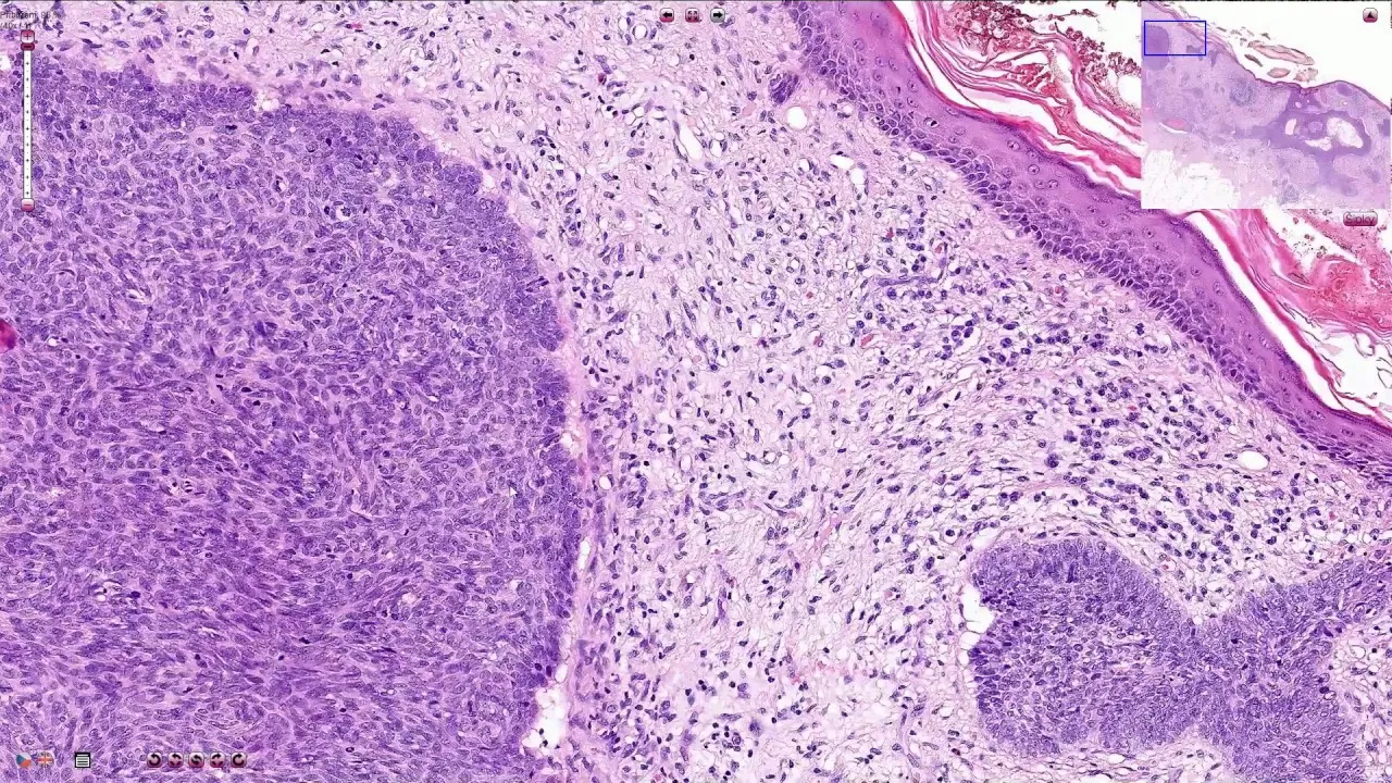
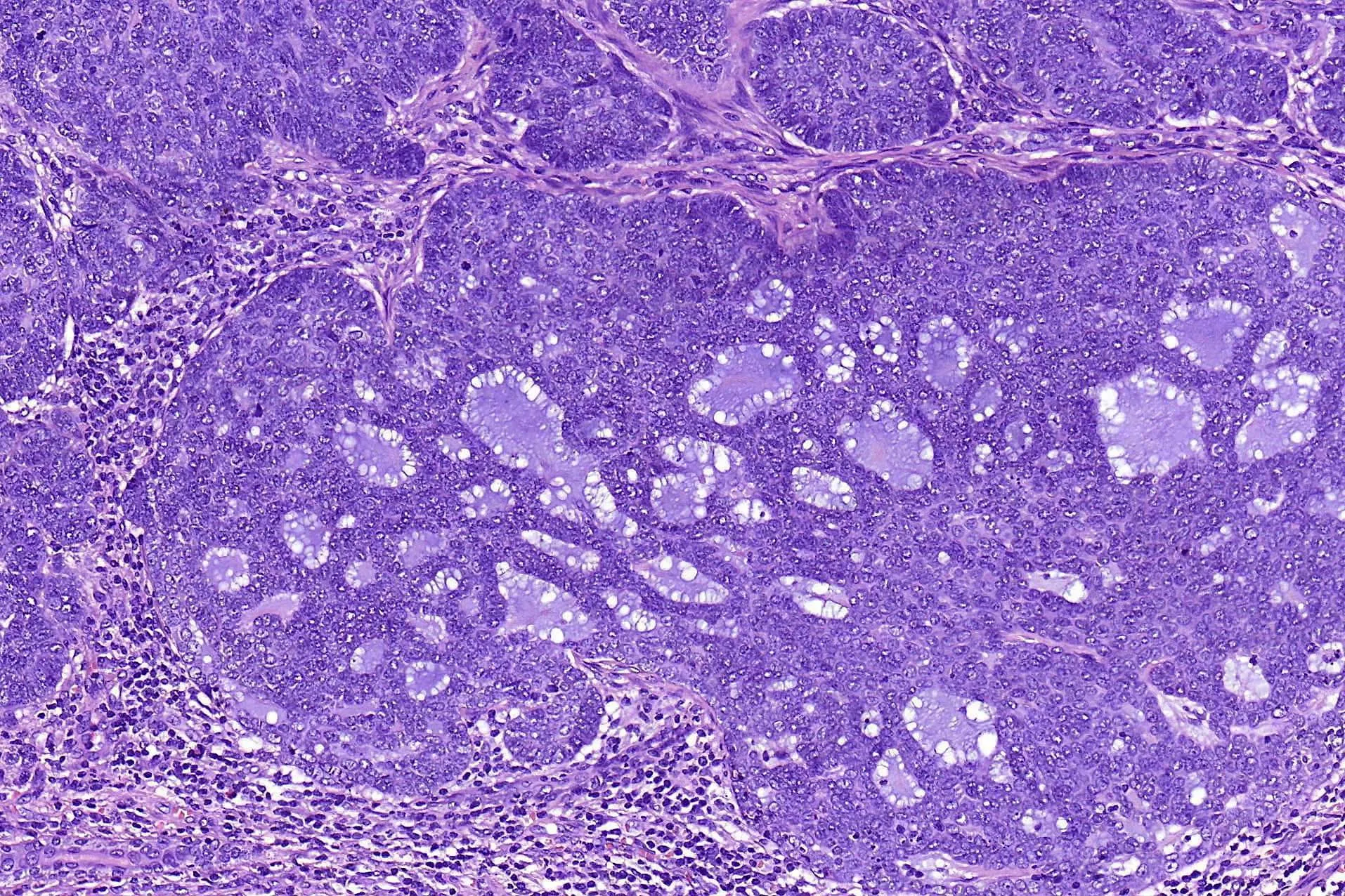
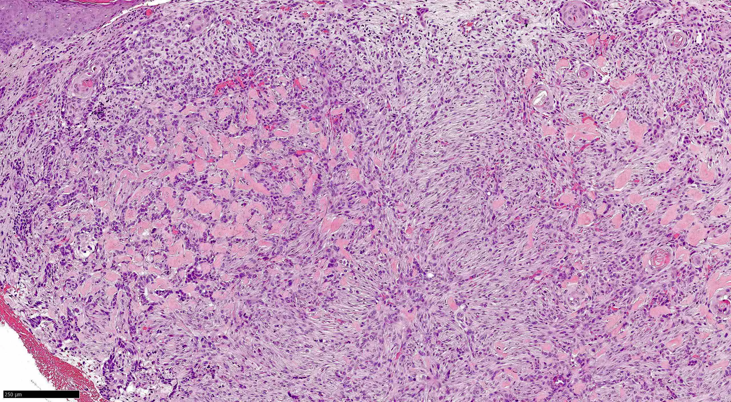
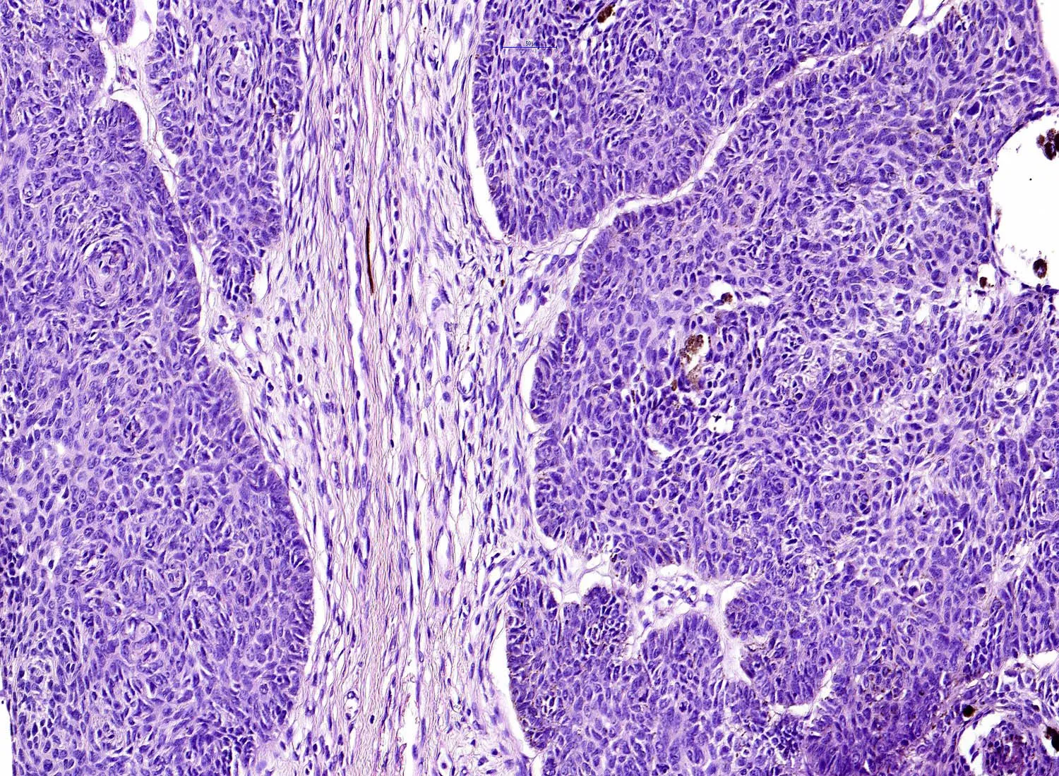
Basal cell tumors in cats are common cutaneous neoplasms that arise from the basal layer of the epidermis. While most are benign and slow-growing, a malignant variant known as basal cell carcinoma can occur. The provided illustration, “Basal cell carcinoma in case 1. The mass consists of a horizontally elongated,” highlights a typical presentation of this cancerous form. Understanding the clinical features, diagnostic approach, and treatment options for feline basal cell tumors helps owners seek timely care. This article explores the nature of these growths, their management, and what to expect during recovery.
What Are Basal Cell Tumors in Cats?
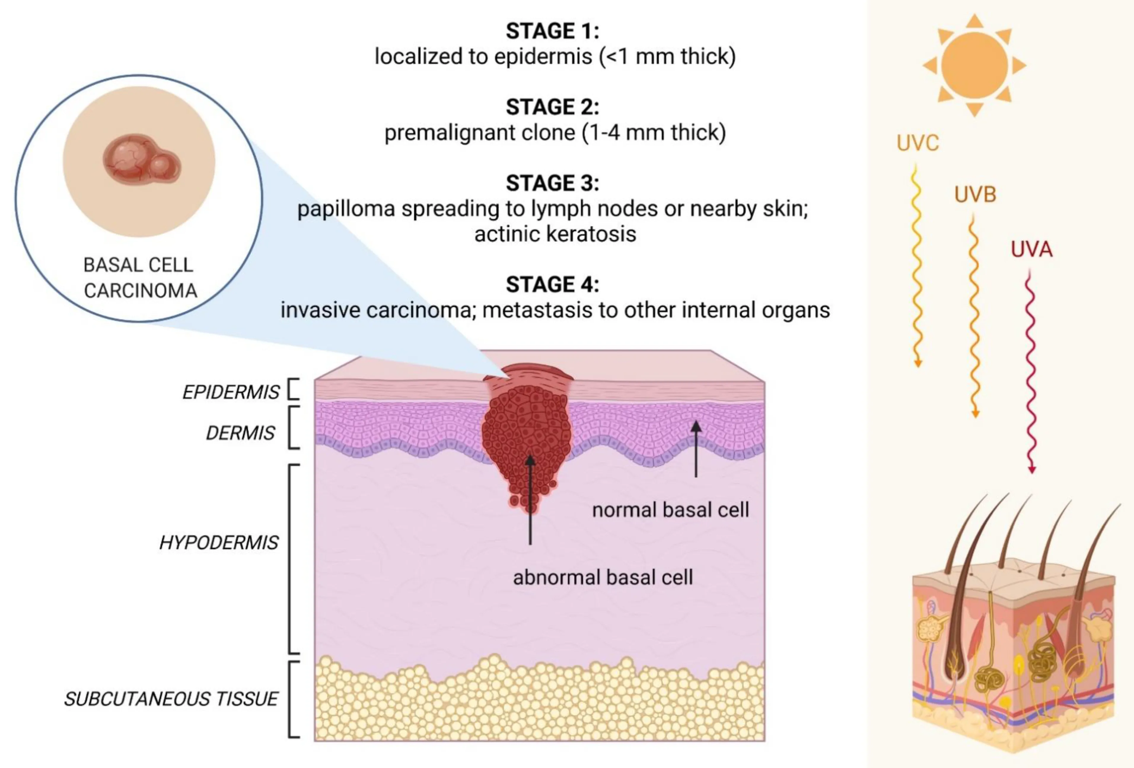
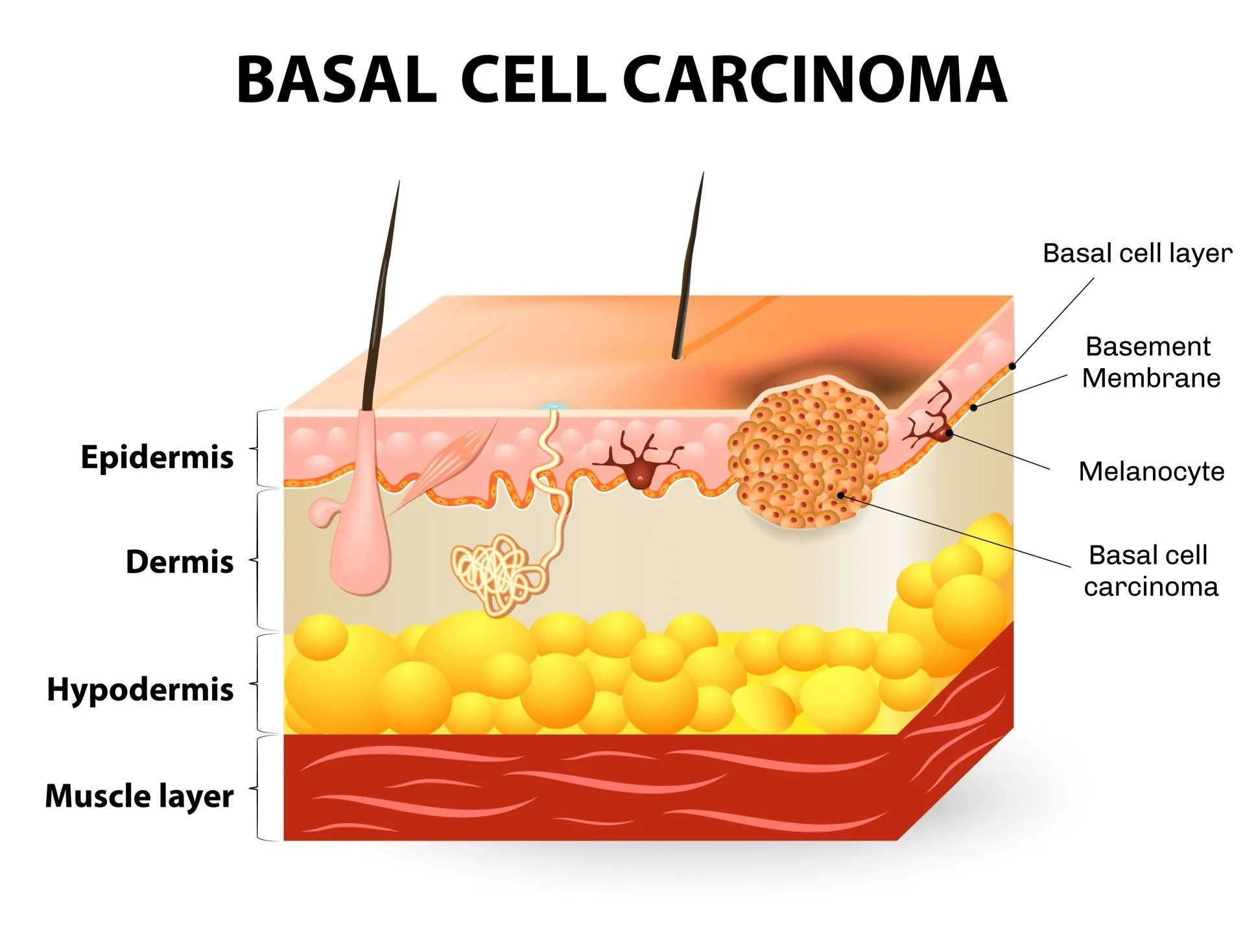
Basal cell tumors are neoplasms originating from the basal cells of the skin’s epidermis. In cats, the vast majority are benign and are classified as basal cell epitheliomas. These growths typically appear as solitary, firm, dome-shaped or plaque-like nodules on the head, neck, or shoulders. According to the Merck Veterinary Manual, basal cell tumors are among the most frequently diagnosed skin neoplasms in cats.
Basal cell carcinoma, the malignant counterpart, is less common but can be locally aggressive. The case of a horizontally elongated mass illustrates the infiltrative pattern of carcinoma. These tumors may ulcerate and invade surrounding tissues but rarely metastasize. Complete surgical excision is essential for a favorable outcome. Understanding common cat diseases helps owners recognize when a skin mass warrants veterinary attention.
Benign vs. Malignant Basal Cell Tumors
Benign basal cell epitheliomas are well-circumscribed, slow-growing, and often pigmented. They rarely cause discomfort unless they become traumatized. In contrast, basal cell carcinoma is characterized by rapid, irregular growth and a tendency to ulcerate. The horizontally elongated mass described in the referenced case is a classic example of a carcinoma that follows tissue planes rather than forming a discrete nodule.
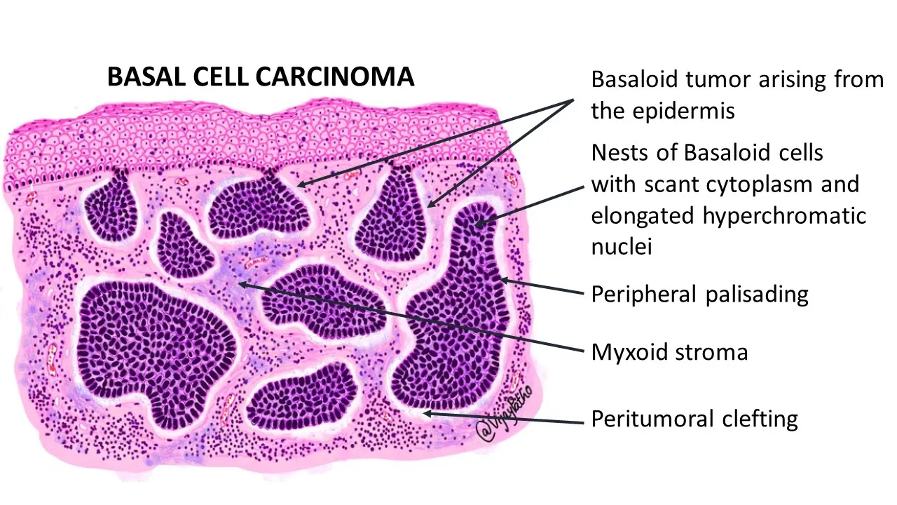
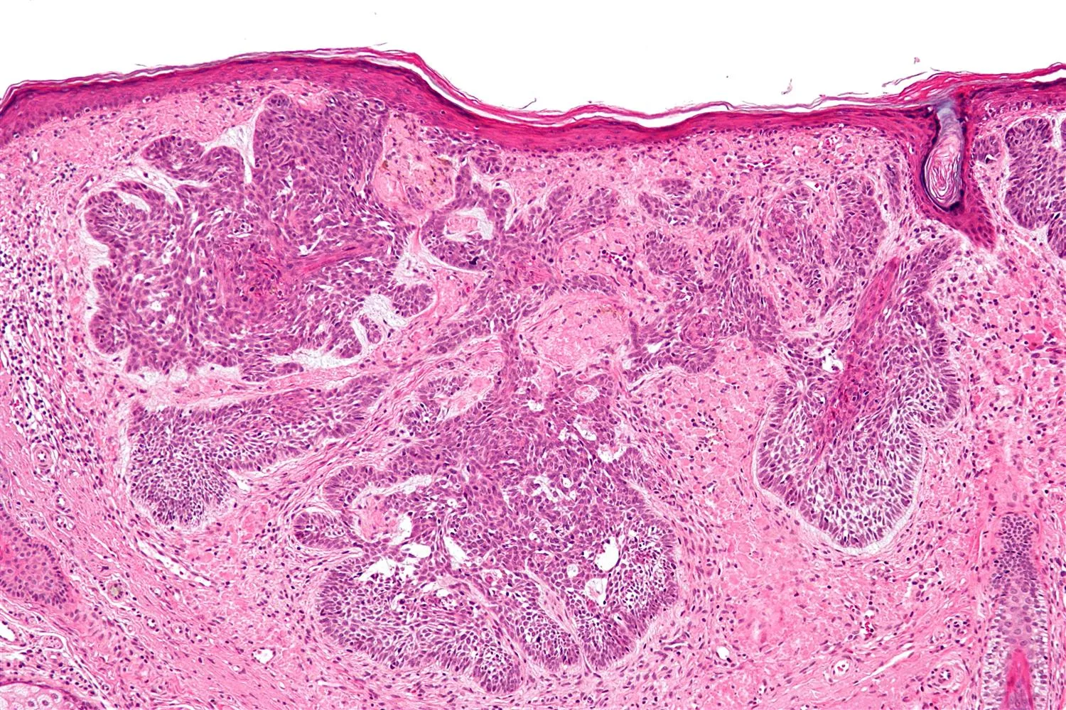
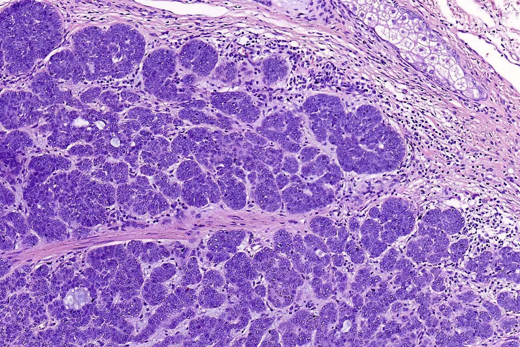
Histologically, benign tumors show orderly basaloid cells with no invasion. Carcinomas exhibit nests of neoplastic cells infiltrating the dermis. While basal cell carcinoma is locally destructive, distant metastasis is exceptionally rare, making aggressive local control the cornerstone of management.
Clinical Signs and Appearance
Basal cell tumors in cats most often present as a single, firm mass on the head, neck, or shoulders. The surface may be smooth or slightly ulcerated, and the color ranges from skin-toned to darkly pigmented. Because the tumors grow slowly, many owners notice them only when they reach a noticeable size. The horizontally elongated configuration, as seen in the case of basal cell carcinoma, occurs when the neoplasm spreads along the dermal layer.
Other signs include hair loss over the mass and, in malignant cases, ulceration, bleeding, or a crusted surface. Cats may lick or scratch the area if it becomes irritated. Unlike inflammatory conditions, basal cell tumors are typically non-painful unless secondary infection develops.
Warning Signs That Require Immediate Veterinary Attention
- Rapid enlargement of a skin mass within days or weeks
- Ulceration, persistent bleeding, or foul-smelling discharge
- Signs of pain, such as vocalization when the area is touched
- Development of multiple new masses in a short period
- Any mass that interferes with movement, eating, or grooming
Diagnosis of Basal Cell Tumors in Cats
A veterinarian begins with a thorough physical examination and a detailed history. Fine needle aspiration is often the first step to obtain cells for cytology. While cytology can suggest a basal cell origin, definitive diagnosis requires a tissue biopsy. Bacterial infections in cats can produce abscesses that mimic tumors, so cytology also helps rule out infection.
Histopathology of an excisional or incisional biopsy reveals the characteristic basaloid cells arranged in nests or cords. For basal cell carcinoma, the pathologist will note invasion into surrounding dermis and possibly mitotic figures. Imaging such as ultrasound or CT may be used to assess local invasion if the tumor is large or fixed to underlying structures.
Treatment Options for Basal Cell Tumors in Cats
Surgical excision is the treatment of choice for both benign and malignant basal cell tumors. For benign epitheliomas, narrow margins are usually curative. Basal cell carcinoma requires wide surgical margins of at least 1–2 cm of normal tissue to minimize the risk of recurrence. The horizontally elongated growth pattern of the carcinoma in the case under discussion underscores the need for careful preoperative planning.
Additional options include cryosurgery for small, superficial benign tumors and radiation therapy for incompletely resected carcinomas. Chemotherapy is rarely needed because of the low metastatic potential. In all cases, the excised tissue should be submitted for histopathology to confirm the diagnosis and evaluate surgical margins.
Home Care and Aftercare Following Tumor Removal
After surgery, the cat must wear an Elizabethan collar to prevent licking or chewing at the incision. The surgical site should be checked daily for swelling, redness, or discharge. Any sutures or staples are typically removed in 10–14 days. The veterinarian may prescribe pain medication and, if necessary, antibiotics. Activity restriction is recommended until the wound heals. Follow-up visits allow the veterinarian to monitor healing and discuss biopsy results.
Distinguishing Basal Cell Tumors from Other Feline Skin Conditions
Several skin conditions can resemble basal cell tumors. Mast cell tumors may appear similar but often cause itching due to histamine release. Injection site sarcomas are more aggressive and occur at vaccine locations. Atopic dermatitis in cats causes pruritus and hair loss, not firm solitary masses. Bartonella in cats can produce papules and lymphadenopathy, but these are inflammatory rather than neoplastic.
Oral basal cell tumors are rare, but bad breath in cats is usually due to dental disease. A mass in the mouth causing halitosis should be biopsied to rule out neoplasia. Sebaceous adenomas and squamous cell carcinomas are other differentials that require histopathology for accurate identification. Cytology and biopsy remain essential for distinguishing these conditions.
Recovery Outlook and Prognosis
Basal cell tumors in cats carry an excellent prognosis when completely excised. Benign epitheliomas are cured by simple removal and rarely recur. Basal cell carcinoma, although locally invasive, has a low metastatic rate. Wide surgical excision typically results in long-term control. Owners should monitor the site for any new growth, but recurrence is uncommon after complete resection. Regular veterinary check-ups help ensure any new skin changes are evaluated promptly.
Healing after surgery is generally straightforward. Cats usually resume normal activity within a few weeks. The cosmetic outcome is good, even for larger excisions. No additional therapy is needed for benign tumors, while carcinomas may require periodic re-examinations. Early detection and treatment provide the best chance for a positive outcome.
Frequently Asked Questions (FAQ)
What is the difference between a basal cell tumor and basal cell carcinoma in cats?
A basal cell tumor is an umbrella term for neoplasms arising from basal cells. Most are benign basal cell epitheliomas. Basal cell carcinoma is the malignant form, characterized by infiltrative growth and local tissue destruction. The carcinoma in the referenced case appeared as a horizontally elongated mass, reflecting its aggressive local spread.
Can basal cell tumors in cats go away on their own?
Basal cell tumors do not resolve spontaneously. Benign tumors may remain stable for years, but they will not shrink without treatment. Malignant carcinomas will continue to grow and invade surrounding tissues. Surgical removal is the only reliable way to eliminate the tumor and prevent potential complications.
How are basal cell tumors diagnosed?
Diagnosis begins with a physical exam and fine needle aspiration for cytology. A definitive diagnosis requires a biopsy and histopathology. The pathologist examines the tissue for basaloid cells and invasion patterns. This confirms whether the tumor is benign or malignant and guides treatment decisions.
Is surgery always necessary for basal cell tumors in cats?
Surgery is the standard treatment for both benign and malignant basal cell tumors. Even benign tumors can become traumatized or interfere with normal function if left untreated. For basal cell carcinoma, wide excision is essential to prevent local recurrence. Non-surgical options like cryosurgery may be appropriate for very small, superficial benign tumors.
What should I watch for after my cat’s tumor removal?
Monitor the incision site daily for redness, swelling, discharge, or gaping. Ensure the cat does not lick or scratch the area by using an Elizabethan collar. Follow the veterinarian’s instructions for pain medication and activity restriction. Attend all follow-up appointments for suture removal and histopathology review.
Basal Cell Tumors in Cats Conclusion
Basal cell tumors in cats are common skin growths that range from benign epitheliomas to locally invasive carcinomas. The horizontally elongated mass described in the case of basal cell carcinoma illustrates the importance of thorough evaluation and surgical planning. Prompt diagnosis and complete excision lead to excellent outcomes. This article is for general informational purposes only. Any cat showing a skin mass or related symptoms should be examined by a licensed veterinarian without delay.