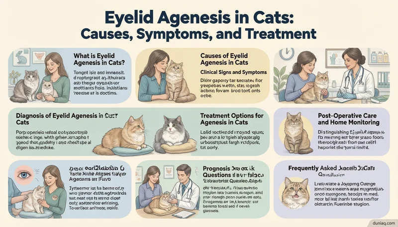
Eyelid agenesis in cats is a rare congenital condition where a segment of the eyelid margin fails to form. The defect usually involves the upper lateral eyelid, leaving the cornea exposed. Affected cats often develop chronic irritation, tearing, and corneal damage. Early recognition and appropriate care are essential to preserve comfort and vision.
Without the protective lid margin, the tear film distribution is disrupted. The cornea becomes vulnerable to injury and scarring. Kittens are born with the anomaly, and clinical signs may appear soon after eye opening. Veterinary ophthalmologic evaluation is critical for a favorable outcome.
What is Eyelid Agenesis in Cats?
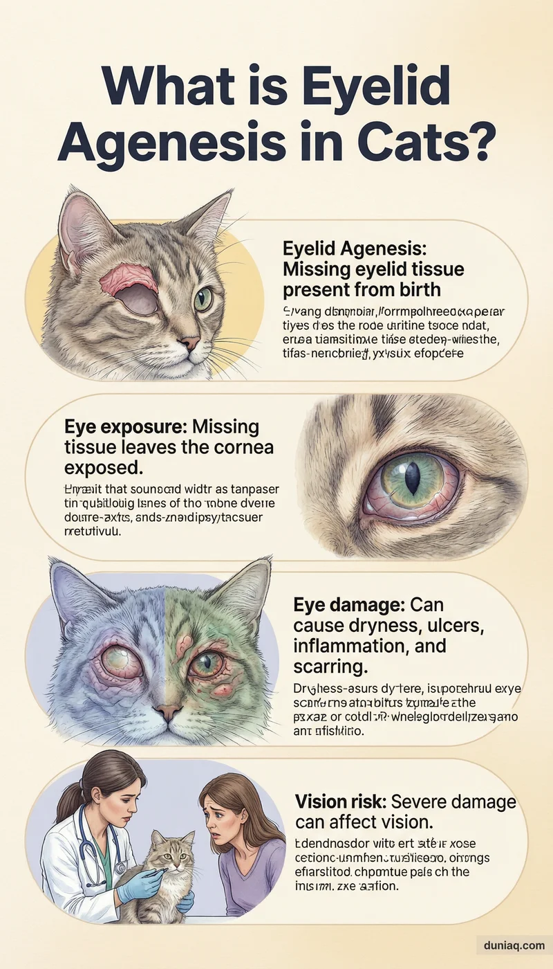
Eyelid agenesis in cats is a developmental anomaly characterized by incomplete formation of eyelid tissue. The defect most commonly affects the lateral portion of the upper eyelid, creating a full‑thickness notch or gap. The missing tissue includes skin, muscle, and conjunctiva. Cats are born with the condition, and it is often bilateral.
This structural failure exposes the cornea to environmental irritants and prevents proper tear film distribution. Without the normal lid margin, the ocular surface becomes dry and prone to ulceration. Chronic inflammation can lead to corneal scarring and vascularization. Prompt recognition helps limit long‑term damage.
Causes of Eyelid Agenesis in Cats
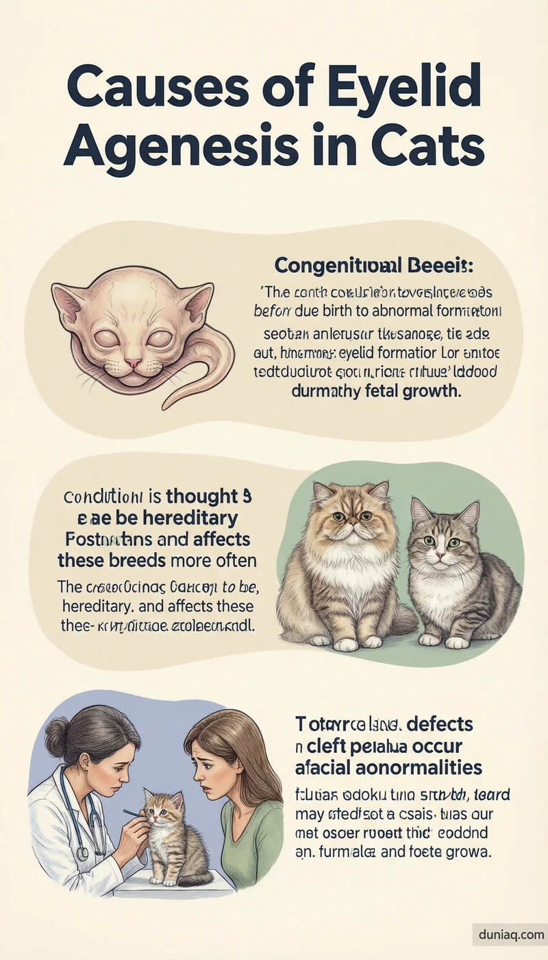
Eyelid agenesis is a congenital defect that arises during embryonic development. The exact cause is not fully understood, but a genetic basis is strongly suspected. Certain breeds, such as domestic shorthairs and Persians, appear overrepresented. It is not an acquired condition and is present from birth.
During fetal growth, the eyelid folds form from surface ectoderm and mesenchyme. Disruption of this process results in agenesis. No specific gene mutation has been definitively identified, but the condition is considered heritable. Environmental factors during pregnancy are not known to play a significant role.
In some cases, eyelid agenesis may occur alongside other craniofacial abnormalities. Cats with eyelid agenesis may also have congenital defects such as cleft palate. This association suggests a broader developmental field defect affecting the face.
Clinical Signs and Symptoms
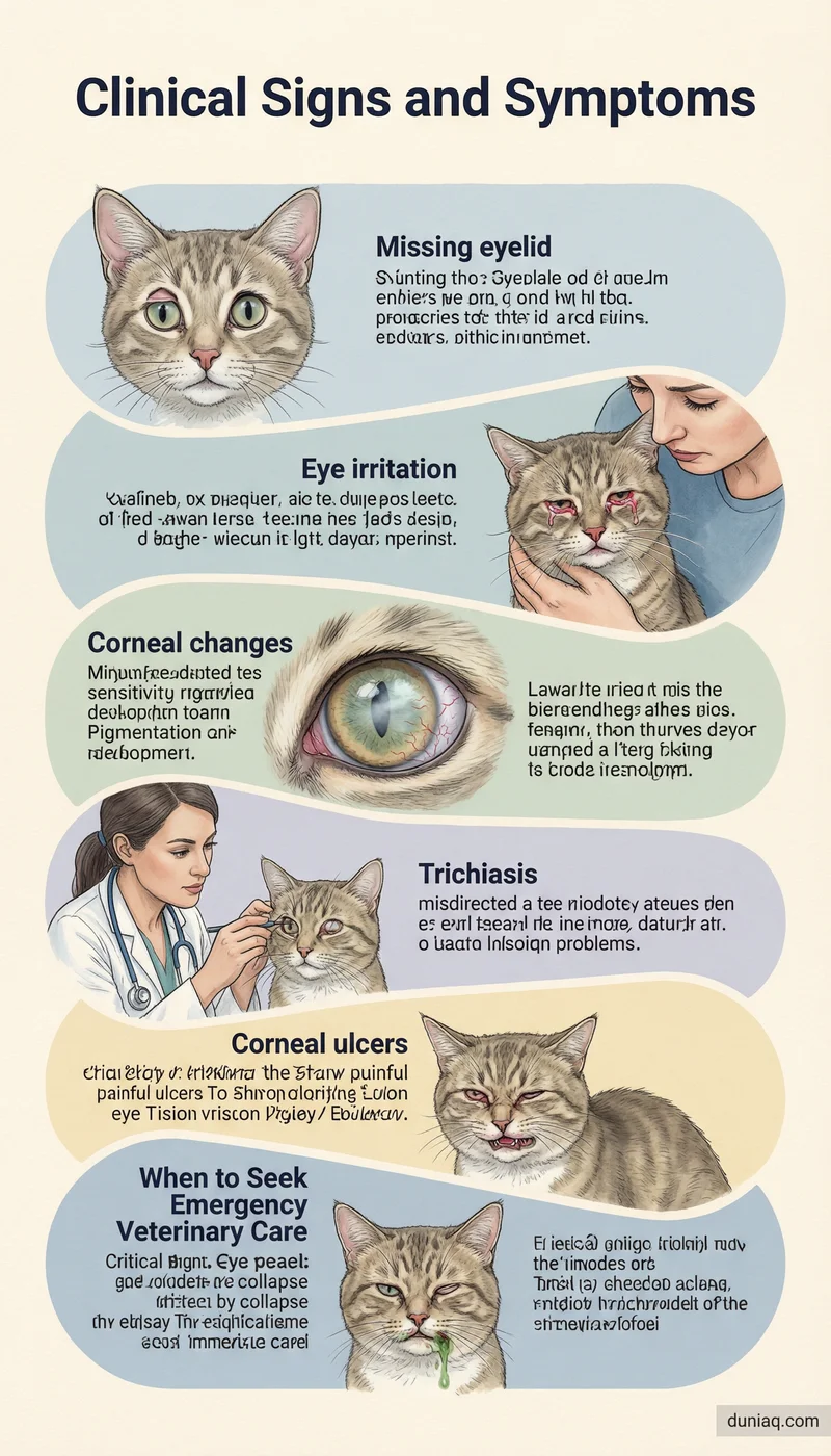
The most noticeable sign of eyelid agenesis in cats is a visible notch or missing segment of the upper eyelid margin. The defect is usually located near the outer corner of the eye. Affected kittens often show excessive tearing, squinting, and light sensitivity. The eye may appear red and irritated.
As the cornea becomes chronically exposed, it develops a dull or hazy appearance. Blood vessels may grow across the corneal surface, and pigmentation can accumulate. Cats may rub at the eye frequently. Secondary corneal ulceration is a common complication, causing pain and discomfort.
Trichiasis, or misdirected hairs from the surrounding skin, frequently rubs against the cornea. These hairs exacerbate irritation and corneal damage. Over time, vision can be impaired if corneal scarring becomes dense. Many cats are presented for evaluation due to persistent ocular discharge and squinting.
When to Seek Emergency Veterinary Care
If a cat with eyelid agenesis suddenly develops severe squinting, cloudiness, or a visible divot on the cornea, urgent veterinary attention is needed. These signs may indicate a deep corneal ulcer or perforation. A perforated globe is a true emergency that can lead to vision loss or loss of the eye.
Any change in pupil shape, collapse of the eye, or a greenish discharge should prompt immediate evaluation. Rapidly worsening pain or refusal to open the eye also warrants emergency care. Early intervention can prevent irreversible damage.
Diagnosis of Eyelid Agenesis in Cats
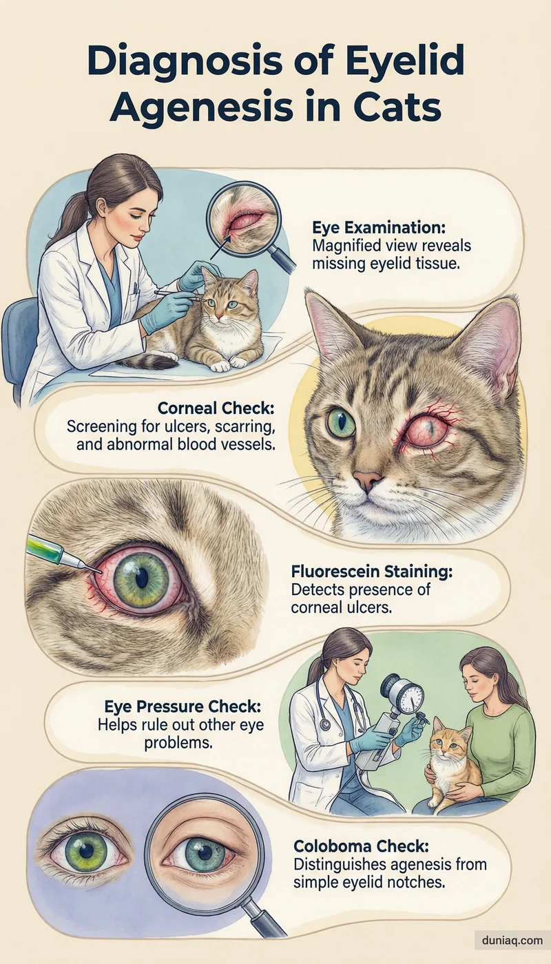
Diagnosis is based on a thorough ophthalmic examination. The veterinarian will inspect the eyelid margins under magnification and good lighting. The characteristic notch or full‑thickness defect in the lateral upper eyelid is usually obvious. The cornea is assessed for ulcers, vascularization, and scarring.
Fluorescein staining is used to detect corneal ulcers and assess the tear film. Intraocular pressure and examination of the anterior chamber help rule out concurrent diseases. Evaluation of the third eyelid and conjunctiva is also performed. The diagnosis of eyelid agenesis in cats is typically straightforward once the defect is visualized.
In some cases, the defect may be subtle and resemble a coloboma. A coloboma is a notch in the eyelid but often involves the margin without full‑thickness tissue loss. Careful examination distinguishes true agenesis from a simple coloboma. Referral to a veterinary ophthalmologist may be recommended for surgical planning.
Treatment Options for Eyelid Agenesis in Cats
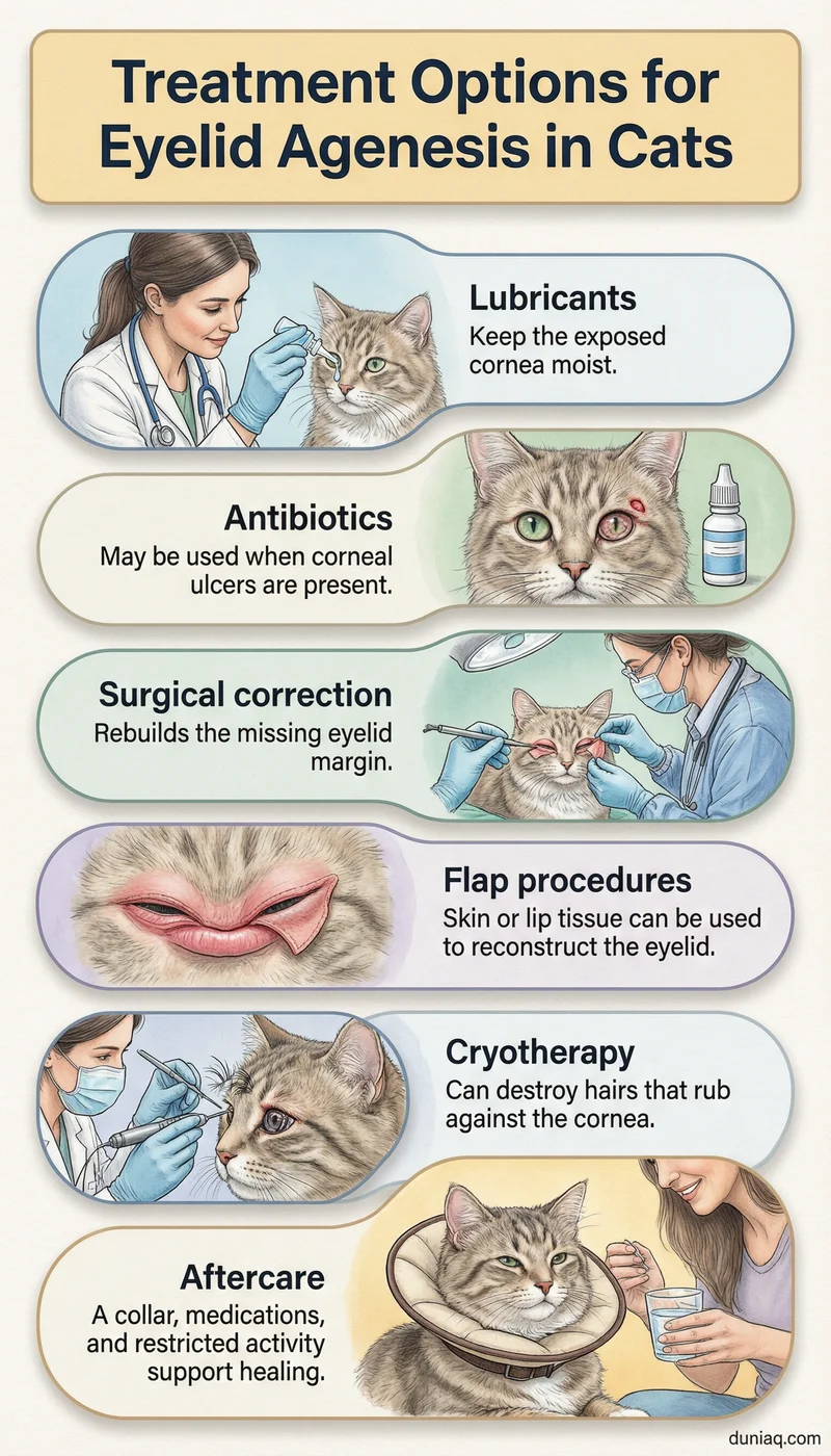
Treatment goals are to protect the cornea, restore eyelid function, and eliminate trichiasis. Medical management alone can provide temporary relief but does not correct the structural defect. Lubricating ointments or artificial tears are often prescribed to keep the cornea moist. Topical antibiotics may be needed if ulceration is present.
Surgical correction is the definitive treatment for eyelid agenesis in cats. The most common procedure involves using a rotational skin flap or a lip‑to‑lid flap to reconstruct the missing eyelid margin. Cryotherapy is often used to destroy aberrant hairs that rub the cornea. The choice of technique depends on the size of the defect.
A frequently used surgical method is the Roberts–Bistner procedure, which employs a mucocutaneous flap. Another option is the lip‑to‑lid flap, where tissue from the lip is transferred to the eyelid. These techniques aim to create a new lid margin that supports normal blinking and tear film distribution. Surgery is best performed by a veterinary ophthalmologist.
Postoperative care includes an Elizabethan collar to prevent self‑trauma, topical medications, and restricted activity. Sutures are typically removed after two weeks. Most cats tolerate the surgery well, and the reconstructed eyelid provides significant improvement in comfort and corneal health.
Post‑Operative Care and Home Monitoring
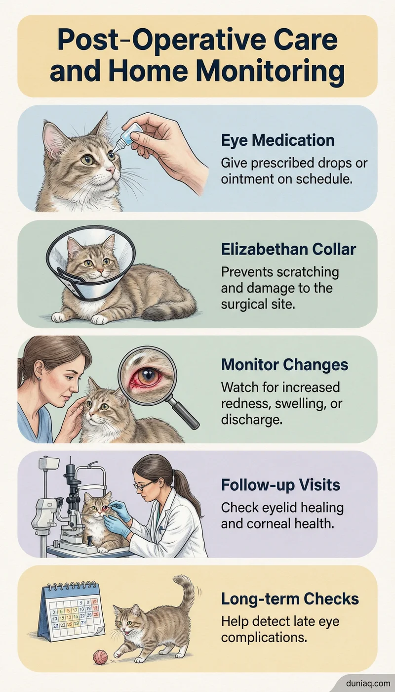
After surgical correction, consistent home care is essential for healing. The owner must administer prescribed eye drops or ointments on schedule. The Elizabethan collar should remain in place until the veterinarian advises removal. Any rubbing or scratching at the eye can compromise the surgical site.
Monitoring for increased redness, swelling, or discharge is important. Mild inflammation is expected, but worsening signs may indicate infection or suture irritation. Follow‑up visits allow the veterinarian to assess wound healing and corneal condition. Long‑term ophthalmic evaluations are often recommended to detect late complications.
Distinguishing Eyelid Agenesis from Other Eye Conditions
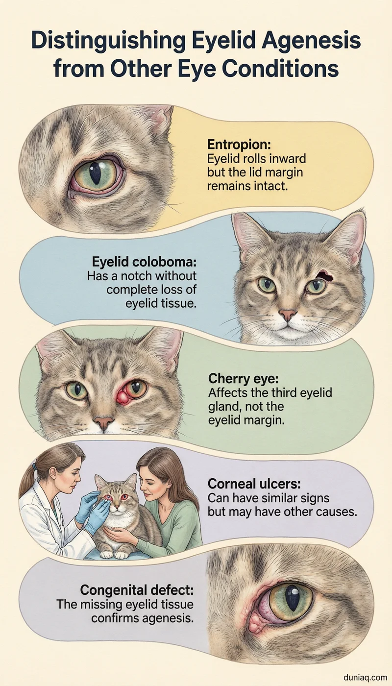
Eyelid agenesis in cats can be confused with other conditions that cause squinting, tearing, and corneal disease. Entropion, where the eyelid rolls inward, also causes corneal irritation but is characterized by an intact lid margin. A thorough examination differentiates these conditions.
Eyelid coloboma is a notch that does not involve full‑thickness tissue loss, whereas agenesis is a true absence of eyelid structures. Cherry eye in cats is a prolapse of the third eyelid gland and does not affect the eyelid margin. These distinctions are important for appropriate treatment planning.
Corneal ulcers from other causes, such as infection or trauma, may mimic secondary signs of agenesis. However, the presence of a congenital eyelid defect confirms the primary diagnosis. A veterinary ophthalmologist can provide a definitive assessment.
Prognosis and Long‑Term Outlook
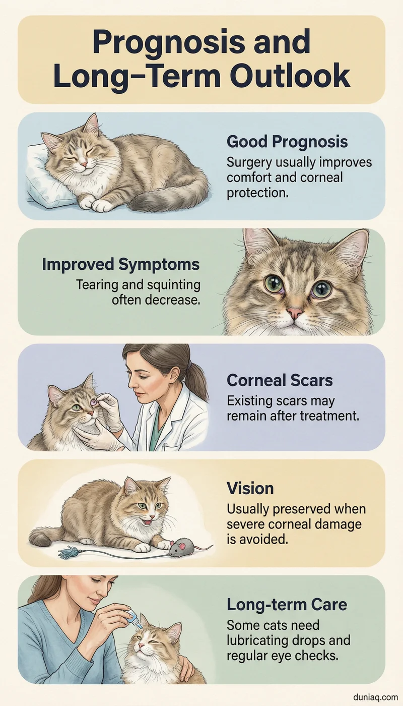
With appropriate surgical intervention, the prognosis for eyelid agenesis in cats is generally good. The reconstructed eyelid significantly improves corneal protection and comfort. Most cats experience reduced tearing, squinting, and corneal vascularization. Lifelong use of lubricating drops may still be needed in some cases.
Corneal scarring present before surgery may not fully resolve, but disease progression is halted. Vision is usually preserved if the cornea was not severely damaged. Regular follow‑up examinations help monitor for recurrence of trichiasis or flap contraction. Early surgical correction offers the best long‑term outcome.
While eyelid agenesis is a structural defect, cats can develop unrelated systemic diseases such as cholangiohepatitis syndrome. Routine wellness care remains important throughout the cat’s life.
Frequently Asked Questions (FAQ) about Eyelid Agenesis in Cats
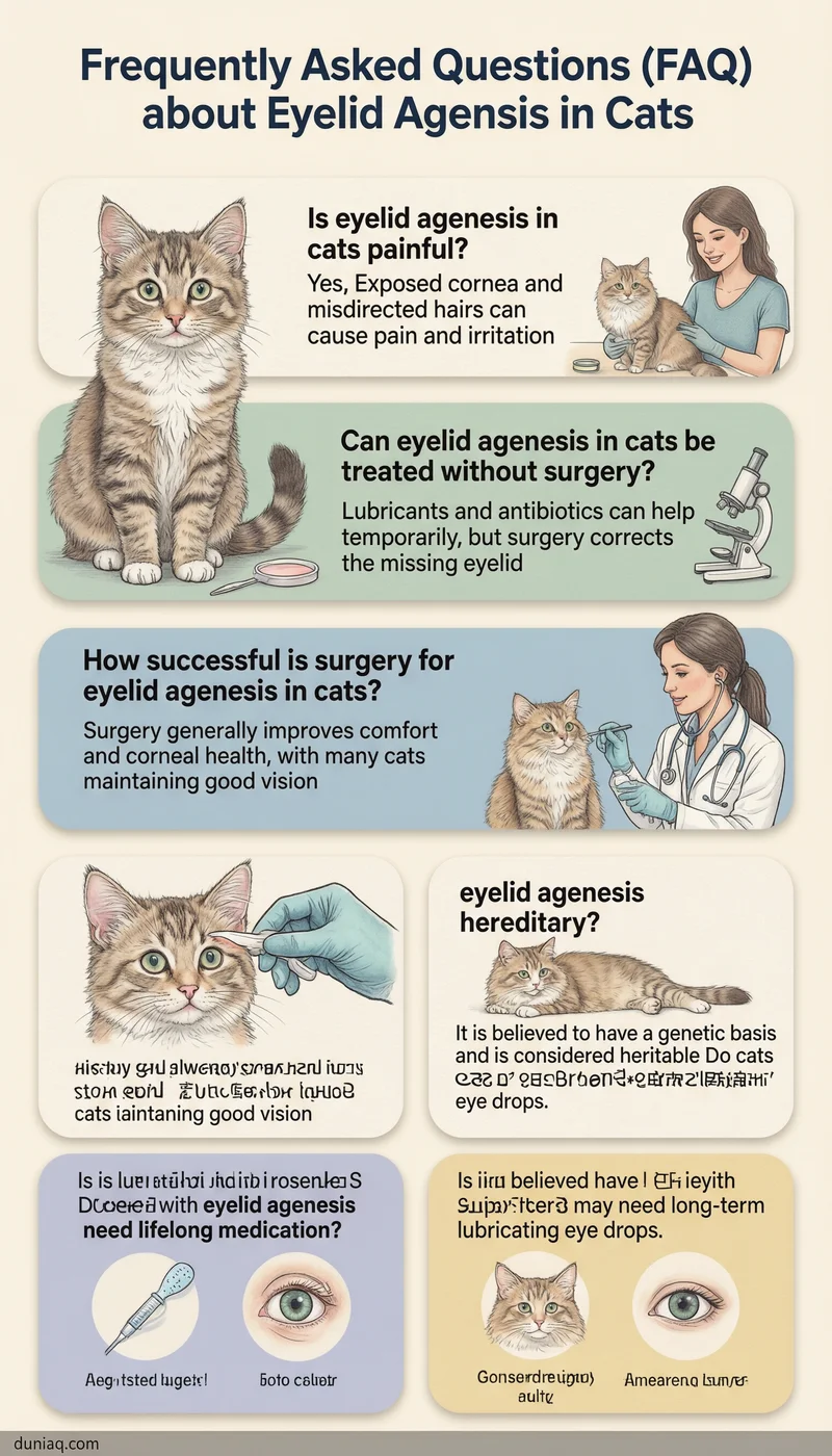
Is eyelid agenesis in cats painful?
Yes, eyelid agenesis in cats is often painful because the exposed cornea becomes dry and irritated. Misdirected hairs rubbing the cornea cause additional discomfort. Surgical correction aims to relieve pain by restoring eyelid function and eliminating trichiasis.
Can eyelid agenesis in cats be treated without surgery?
Medical therapy with lubricants and antibiotics can manage symptoms temporarily but cannot correct the missing eyelid margin. Surgery is the definitive treatment to protect the cornea and prevent long‑term damage. Without surgery, chronic corneal disease is likely to progress.
How successful is surgery for eyelid agenesis in cats?
Surgical reconstruction is generally successful in improving comfort and corneal health. Most cats show a significant reduction in tearing and squinting. The outcome depends on the size of the defect and the expertise of the surgeon, but many cats maintain good vision after surgery.
Is eyelid agenesis hereditary?
Eyelid agenesis is believed to have a genetic basis, as it is a congenital defect present at birth. Affected cats should not be used for breeding. The exact mode of inheritance is not fully understood, but it is considered heritable.
Do cats with eyelid agenesis need lifelong medication?
Some cats may require long‑term use of lubricating eye drops even after successful surgery. The need depends on the degree of corneal health and tear film stability. Regular veterinary checkups help determine if ongoing medication is necessary.
Eyelid Agenesis in Cats Conclusion
Eyelid agenesis in cats is a congenital condition that threatens corneal health and vision. Early recognition, thorough ophthalmic evaluation, and timely surgical reconstruction are key to a comfortable outcome. With proper care, most affected cats can enjoy a good quality of life. This article is for general informational purposes only. Any cat showing eye symptoms should be evaluated by a licensed veterinarian. Veterinary ophthalmologists, such as those certified by the American College of Veterinary Ophthalmologists, provide specialized care for complex eyelid disorders.