Iris melanosis in cats is a pigment change affecting the colored portion of the eye, appearing as flat brown or black spots that gradually spread across the iris surface. Many owners first notice these freckle-like marks during routine play or grooming, often in middle-aged or older cats.
While the majority of these pigment changes remain benign throughout a cat’s life, a small subset can transform into diffuse iris melanoma, an aggressive intraocular cancer. Because appearance alone cannot reliably separate the two, veterinary monitoring matters. Understanding what iris melanosis in cats looks like, how it progresses, and when it warrants concern helps owners respond appropriately.
What is Iris Melanosis in Cats?
Iris melanosis in cats describes an increased accumulation of melanin-producing cells within the surface layer of the iris. The result is one or more flat, sharply defined pigmented patches that do not thicken or distort the iris architecture.
These lesions sit on the anterior iris border, remaining level with surrounding tissue. Pupil shape, movement, and eye pressure stay normal. The condition itself causes no pain, no vision loss, and no inflammation, which distinguishes benign melanosis from malignant transformation.
How Iris Melanosis Differs From Normal Iris Freckles
Cats can be born with small pigment spots called iris nevi, which behave much like human freckles. These remain static in size, number, and color for years. Iris melanosis in cats, by contrast, is progressive: spots enlarge, multiply, and often coalesce into broader pigmented sheets over months to years.
The distinction matters clinically. Static nevi rarely require anything beyond documentation. Progressive pigment spread requires scheduled reevaluation because progression is one of the features shared with early diffuse iris melanoma.
Comparison of Common Iris Pigment Changes
| Feature | Iris Nevus (Freckle) | Iris Melanosis | Diffuse Iris Melanoma |
|---|---|---|---|
| Surface texture | Flat | Flat | Raised, velvety, or thickened |
| Progression | Static | Slow, gradual spread | Spreading, may accelerate |
| Pupil shape | Normal | Normal | May become irregular or sluggish |
| Eye pressure | Normal | Normal | May rise (secondary glaucoma) |
| Comfort | Painless | Painless | May become painful |
Causes of Iris Melanosis in Cats
The underlying trigger for iris melanosis in cats has not been definitively established in veterinary literature. Current understanding centers on a localized proliferation of melanocytes—the pigment cells normally present in iris tissue—without the cellular disorganization seen in cancer.
Several contributing factors have been proposed or observed clinically:
- Age: Pigment changes appear most often in middle-aged and senior cats, suggesting cumulative cellular change over time.
- Genetic predisposition: Some cats appear more prone to melanocyte proliferation, though no specific inherited gene has been confirmed.
- Chronic ocular inflammation: Prior or ongoing uveitis has been associated with pigment disturbance in some cases, though causation remains unproven.
- Ultraviolet exposure: Suspected as a contributor by analogy with skin melanocytes, but not established in feline eyes.
Importantly, iris melanosis in cats is not caused by diet, injury from ordinary play, or infectious agents. Owners should not attribute the change to something they did or failed to do.
Breed and Coat Color Considerations
No breed has been reliably identified as high-risk. Cats of any coat color and any eye color—green, gold, copper, or blue—can develop pigment spots. Lighter irises simply make the spots more visible earlier, which can create the false impression that certain cats are more affected.
Symptoms and Signs of Iris Melanosis in Cats
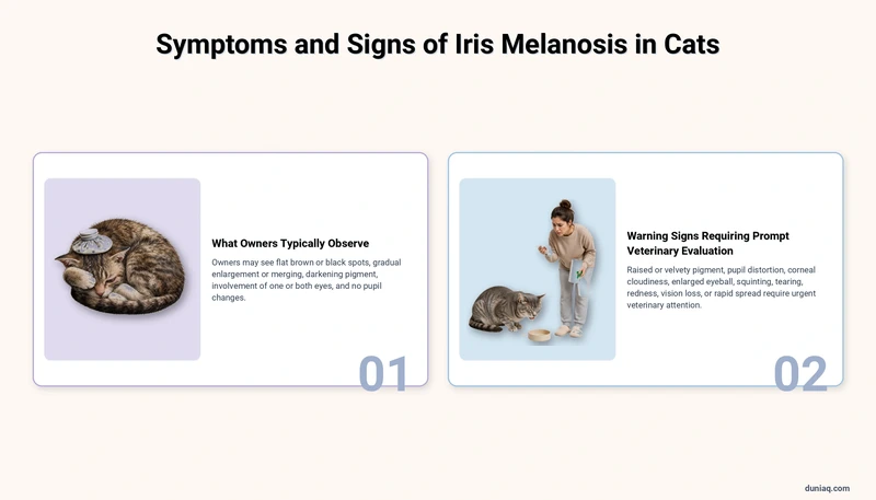
The primary sign is visual and painless. Most cats behave entirely normally, and the change is detected by an observant owner or during a wellness exam. There is no discharge, squinting, or rubbing associated with uncomplicated iris melanosis in cats.
What Owners Typically Observe
- One or several flat brown or black spots on the colored part of the eye
- Spots that appear like freckles, sometimes irregularly shaped
- Gradual enlargement or merging of spots over months
- Pigment sometimes darkening from light tan to deep brown
- Involvement of one eye, or occasionally both eyes independently
- No change in pupil size, shape, or responsiveness
Because the process is slow, comparing photographs taken months apart is far more revealing than day-to-day observation. Owners are encouraged to photograph the eye under consistent, indirect lighting.
Warning Signs Requiring Prompt Veterinary Evaluation
Certain findings suggest the pigment may no longer be behaving benignly, or that a separate ocular disease is present. These warrant same-day or urgent attention:
- Pigmented area that looks raised, bumpy, or velvety rather than flat
- Pupil that becomes distorted, oval, or slow to constrict in light
- Cloudiness of the cornea or a visibly enlarged eyeball
- Squinting, tearing, light sensitivity, or pawing at the eye
- Redness of the white of the eye or visible blood in the front chamber
- Apparent vision loss, bumping into objects, or reluctance to move in dim light
- Rapid pigment spread over a few weeks rather than months
Sudden ocular pain in cats can accompany several serious conditions. As with other abrupt neurological or systemic changes such as seizure activity in cats, prompt professional assessment is the safest course.
Diagnosis of Iris Melanosis in Cats
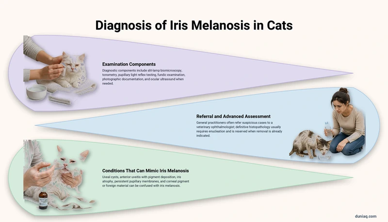
Diagnosis rests on careful ophthalmic examination and, critically, on documented change over time. No single in-office test can definitively separate benign melanosis from early diffuse iris melanoma, which is why serial monitoring forms the backbone of the diagnostic approach.
Examination Components
- Slit-lamp biomicroscopy: Provides magnified, illuminated assessment of whether the pigment is flat or elevated and whether iris tissue architecture is intact.
- Tonometry: Measures intraocular pressure. Elevated pressure raises concern for melanoma cells obstructing fluid drainage.
- Pupillary light reflex testing: Confirms the iris muscle is functioning normally.
- Fundic examination: Evaluates the retina and optic nerve for secondary changes.
- Photographic documentation: Establishes an objective baseline for comparison at future visits.
- Ocular ultrasound: Used when the pigment is dense or the eye is cloudy, to assess for masses behind the iris.
Referral and Advanced Assessment
General practitioners frequently refer suspicious cases to a board-certified veterinary ophthalmologist. Specialists may recommend high-resolution imaging or, in select cases, sampling of aqueous fluid. Definitive histopathology requires tissue, which in practice usually means examining the eye after enucleation—so it is reserved for cases where removal is already indicated.
The American College of Veterinary Ophthalmologists maintains a directory of certified specialists for owners seeking referral.
Conditions That Can Mimic Iris Melanosis
Several other ocular findings are confused with iris melanosis in cats:
- Uveal cysts: Fluid-filled, often free-floating, round and translucent when transilluminated.
- Anterior uveitis with pigment deposition: Accompanied by redness, pain, and flare in the anterior chamber.
- Iris atrophy: Thinning of iris tissue in older cats, which can create dark-appearing areas that are actually holes.
- Persistent pupillary membranes: Congenital strands of tissue, present from kittenhood and non-progressive.
- Corneal pigment or foreign material: Located on the eye surface rather than within the iris.
Treatment Options for Iris Melanosis in Cats
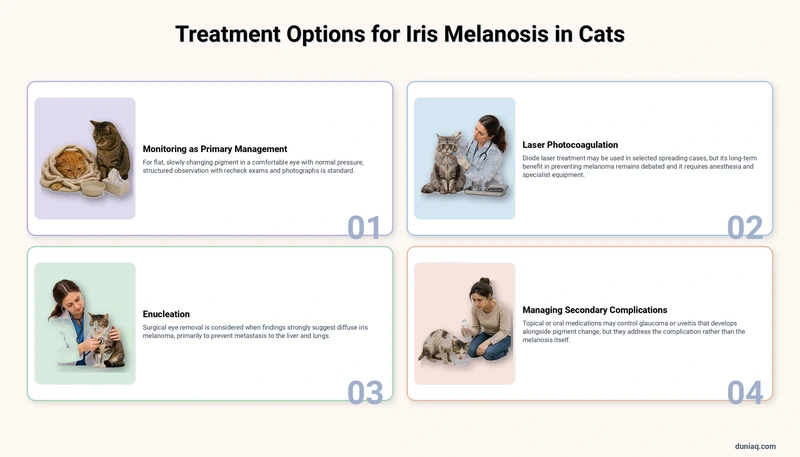
Treatment depends entirely on whether the pigment is judged benign and stable, or progressive and suspicious. There is no medication that removes or reverses iris pigment.
Monitoring as Primary Management
For flat, slowly changing pigment in a comfortable eye with normal pressure, structured observation is the standard approach. This typically involves recheck examinations at intervals determined by the veterinarian, with photographs compared at each visit. Many cats live their entire lives without the condition progressing beyond cosmetic change.
Laser Photocoagulation
In selected cases where pigment is spreading but the eye remains otherwise healthy, a veterinary ophthalmologist may use diode laser treatment to destroy pigmented cells on the iris surface. This is not universally recommended and its long-term benefit in preventing melanoma remains debated within the profession. It requires general anesthesia and specialist equipment.
Enucleation
Surgical removal of the eye is considered when findings strongly suggest diffuse iris melanoma—raised pigment, distorted pupil, secondary glaucoma, or rapid progression. Because this tumor can metastasize to the liver and lungs, removing the eye before spread occurs is the primary strategy for protecting the cat’s overall health.
Cats adapt remarkably well to vision in one eye. Most resume normal navigation, jumping, and hunting behaviors within weeks. The removed eye is submitted for histopathology, which confirms the diagnosis and provides information about tumor invasion.
Managing Secondary Complications
If glaucoma or uveitis develops alongside pigment change, topical or oral medications may be prescribed to control pressure and inflammation. These address the complication rather than the melanosis itself.
Home Care and Ongoing Monitoring
Owners play a central role in tracking iris melanosis in cats between veterinary visits. Practical steps include:
- Photographing each eye monthly in consistent lighting, without flash directed into the eye
- Keeping images dated in a single folder for easy comparison
- Observing pupil symmetry in both bright and dim conditions
- Noting any change in behavior, such as hesitancy on stairs or altered depth perception
- Attending all scheduled recheck appointments even when the eye looks unchanged
After Laser Treatment or Enucleation
Following any ocular procedure, general aftercare usually involves administering prescribed medications exactly as directed, using an Elizabethan collar to prevent self-trauma, restricting jumping and rough play during healing, and keeping the surgical site clean and dry. Follow-up visits allow the veterinary team to assess healing and remove sutures when appropriate.
Some cats resist eye medications. Working calmly and pairing handling with rewards helps; stress-related behaviors like compulsive fur pulling can emerge in cats subjected to repeated unpleasant handling, so gentle technique matters.
Prognosis and What Owners Can Expect
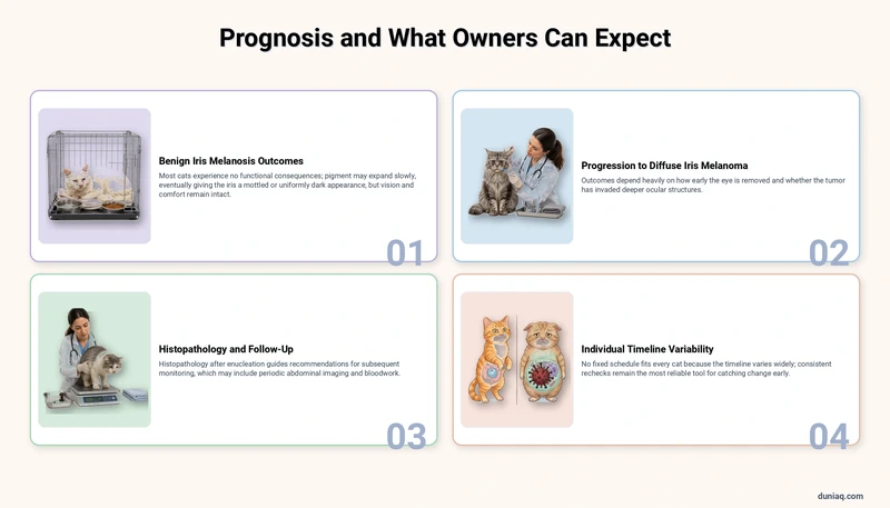
Most cats diagnosed with benign iris melanosis experience no functional consequences. The pigment may continue expanding slowly, eventually giving the iris a mottled or uniformly dark appearance, yet vision and comfort remain intact.
When progression to diffuse iris melanoma occurs, outcomes depend heavily on how early the eye is removed and whether the tumor has invaded deeper ocular structures. Histopathology after enucleation guides the veterinarian’s recommendations for subsequent monitoring, which may include periodic abdominal imaging and bloodwork.
Because the timeline varies widely between individuals, no fixed schedule fits every cat. Consistent rechecks remain the most reliable tool for catching change early.
Prevention and Risk Reduction
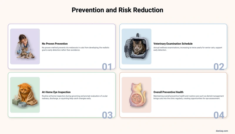
No proven method prevents iris melanosis in cats from developing. The realistic goal is early detection rather than avoidance. Owners can support this through:
- Annual wellness examinations, increasing to twice yearly for senior cats
- Routine at-home inspection of the eyes during grooming sessions
- Prompt evaluation of any ocular redness, discharge, or squinting
- Maintaining overall preventive health, since systemic illness can complicate ocular disease
General feline health awareness supports early detection across many conditions. Familiarity with common feline health conditions helps owners recognize when something has genuinely changed. Routine care such as managing dental plaque and tartar buildup also brings cats into the clinic regularly, creating natural opportunities for eye assessment.
Additional peer-reviewed guidance on feline ocular tumors is available through the Merck Veterinary Manual.
Frequently Asked Questions (FAQ) about Iris Melanosis in Cats
Is iris melanosis in cats painful?
Benign iris melanosis causes no pain. The pigment sits flat within the iris and does not irritate surrounding tissue. If your cat squints, tears, or avoids light, a different or additional problem is likely present and needs veterinary assessment.
Can iris melanosis in cats turn into cancer?
A minority of cases progress to diffuse iris melanoma. There is currently no test that predicts which cats will progress, which is why veterinarians recommend photographic documentation and scheduled reexaminations rather than a single one-time assessment.
Does iris melanosis affect my cat’s vision?
Flat pigment on the iris surface does not block light entering the pupil, so vision is unaffected. Vision changes suggest complications such as glaucoma, uveitis, or a mass lesion, all of which require prompt examination.
How quickly does iris melanosis in cats spread?
Progression is typically measured in months to years and is often imperceptible day to day. Pigment that visibly expands over a few weeks is considered atypical and should prompt an earlier recheck with your veterinarian.
Should both of my cat’s eyes be examined?
Yes. Iris melanosis in cats can affect one or both eyes, sometimes at different rates. Comparing the two eyes at each visit also helps the veterinarian detect subtle asymmetry in pupil function or pigment distribution.
Iris Melanosis in Cats Conclusion
Iris melanosis in cats is usually a benign, painless pigment change that requires observation rather than intervention. The key responsibility for owners is consistent documentation and attending recommended rechecks, since progression to melanoma cannot be ruled out by appearance alone. This article provides general information only. Any cat showing eye changes or discomfort should be examined by a licensed veterinarian.