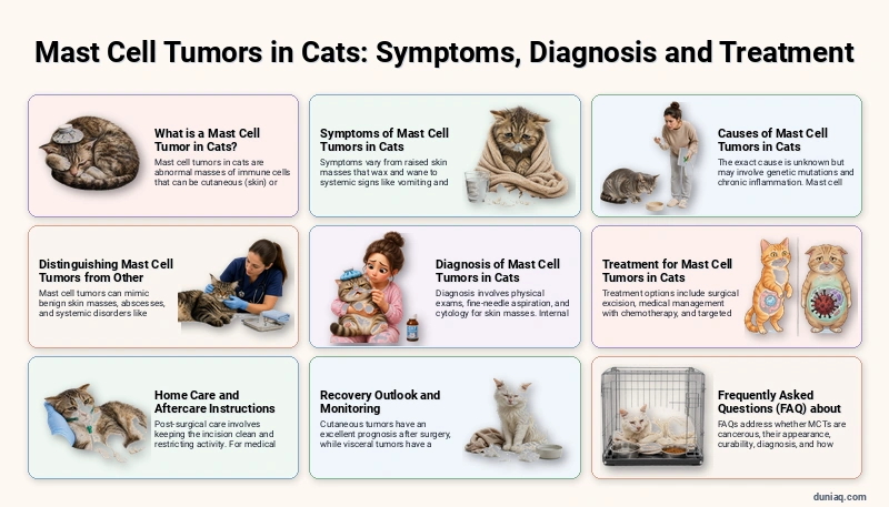
Mast cell tumors in Cats (MCTs) are neoplasms arising from mast cells, immune cells packed with histamine and heparin. In cats, they commonly develop on the skin, especially the head and neck, but can also affect internal organs such as the spleen and intestines. Cutaneous mast cell tumors may appear as firm nodules that fluctuate in size due to degranulation.
Feline mast cell tumors exhibit variable behavior; many skin tumors are benign and cured by excision, while visceral forms are aggressive. Symptoms depend on tumor location and histamine release, causing redness, itching, or gastrointestinal upset. Recognizing early signs enables prompt veterinary intervention and better outcomes.
What is a Mast Cell Tumor in Cats?
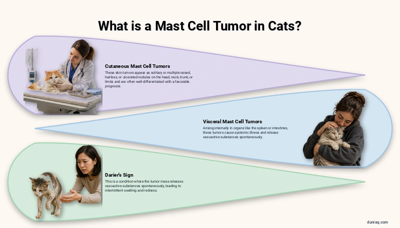
Mast cells are normal immune cells residing in connective tissues, ready to respond to allergens and parasites by releasing histamine, proteases, and cytokines. When these cells undergo neoplastic transformation, they multiply uncontrollably, forming a mass that may release vasoactive substances spontaneously, leading to intermittent swelling and redness known as Darier’s sign. In cats, mast cell tumors are categorized into cutaneous (skin) and visceral (internal) forms.
Cutaneous mast cell tumors typically present as solitary or multiple raised nodules on the head, neck, trunk, or limbs. They can be hairless, erythematous, and ulcerated. Visceral mast cell tumors often arise in the spleen (splenic mast cell tumor) or intestines, causing systemic illness. Histologically, feline cutaneous MCTs are often well-differentiated, which correlates with a more favorable prognosis compared to dogs.
Symptoms of Mast Cell Tumors in Cats
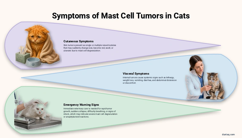
The presentation of mast cell tumors in cats varies widely. Cutaneous forms typically manifest as a single, raised mass or multiple nodules. These growths may appear suddenly, change size, or become red and swollen after manipulation because mast cells degranulate. Owners often notice a lump that seems to wax and wane, sometimes accompanied by ulceration and bleeding. Mild pruritus is possible.
Visceral mast cell tumors usually cause systemic signs. Cats may show lethargy, weight loss, vomiting, diarrhea, and decreased appetite. Splenic tumors can lead to abdominal distension or discomfort. Intestinal tumors may produce chronic gastrointestinal signs similar to inflammatory conditions. Episodic redness and swelling around the tumor site can occur due to histamine release, a helpful clue during a physical exam.
Emergency Warning Signs
Certain symptoms require immediate veterinary attention. Rapid tumor growth, sudden collapse, difficulty breathing, or severe vomiting and diarrhea could indicate mast cell degranulation syndrome or anaphylactoid reactions. If a cat appears weak, has pale gums, or shows signs of shock after handling a mass, this is a medical emergency. Prompt intervention can stabilize life-threatening histamine release.
Causes of Mast Cell Tumors in Cats
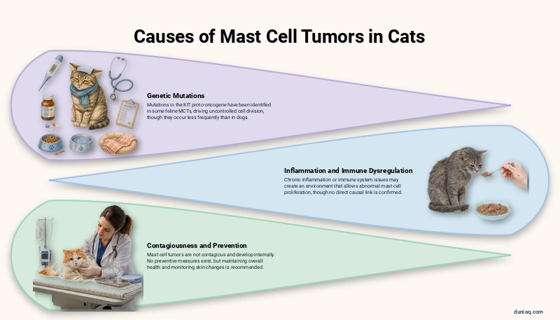
The exact cause of mast cell tumors in cats remains unknown. Genetic mutations in the KIT proto-oncogene have been identified in some feline MCTs, leading to uncontrolled cell division. Unlike dogs, cats less frequently carry specific KIT mutations, but similar pathways may drive tumor growth. Ongoing research aims to clarify the molecular triggers behind spontaneous mast cell neoplasia in this species.
Chronic inflammation or immune dysregulation may create a permissive environment for abnormal mast cell proliferation. Certain feline diseases, including viral infections and chronic inflammatory disorders, have been proposed as potential contributing factors, but no direct causal link has been confirmed. Environmental triggers such as chemical exposure or UV radiation are not established risk factors for cats. A genetic predisposition in some breeds has been suggested but remains unproven.
Mast cell tumors are not contagious. They develop internally from the cat’s own cells. While no preventive measures exist, maintaining overall health and monitoring for any skin changes is the best approach. A solid understanding of common cat diseases helps owners recognize when a veterinary visit is warranted.
Distinguishing Mast Cell Tumors from Other Conditions

Mast cell tumors can mimic a variety of feline skin masses. Benign fatty tumors (lipomas in cats) are soft, movable, and non‑ulcerated, which contrasts with the firmer, often erythematous appearance of cutaneous MCTs. Abscesses and insect‑bite reactions may also cause red, swollen lumps but usually resolve quickly with appropriate treatment and lack the persistent nature of neoplastic growth.
Visceral mast cell tumors produce vomiting, diarrhea, and weight loss that might be confused with inflammatory bowel disease, intestinal lymphoma, or systemic disorders like kidney failure. The waxing‑and‑waning mass near the throat could raise suspicion of laryngitis in cats if voice changes occur. Intestinal forms can disrupt the gut barrier, creating signs reminiscent of leaky gut syndrome, making accurate diagnostic workup essential.
Diagnosis of Mast Cell Tumors in Cats
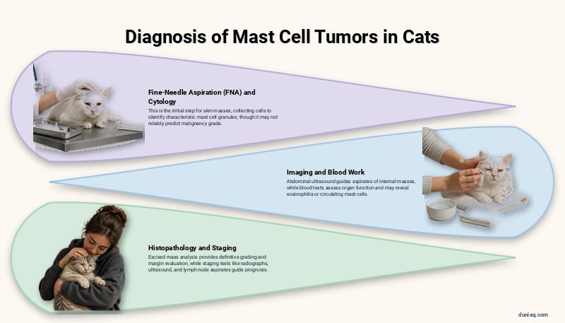
Veterinary diagnosis begins with a thorough physical examination and history. For skin masses, fine‑needle aspiration (FNA) with cytology is the initial step. A small‑gauge needle collects cells, which are stained and examined under a microscope. Mast cells are readily identified by their characteristic cytoplasmic granules. Cytology can confirm mast cell tumor but may not reliably predict malignancy grade in cats.
If the tumor is located internally, abdominal ultrasound guides fine‑needle aspiration of spleen or intestinal masses. Blood work, including a complete blood count and serum biochemistry, assesses organ function and may reveal mast cell‑related abnormalities such as eosinophilia. In some cases, the buffy coat preparation detects circulating mast cells, though this test has limited sensitivity. Histopathology of an excised mass provides definitive grading and margin evaluation.
Staging tests for suspected aggressive tumors include thoracic radiographs, abdominal ultrasound, and lymph node aspirates. A bone marrow aspirate is reserved for suspected systemic mastocytosis. Advanced imaging like computed tomography (CT) may be recommended for extensive visceral involvement. Accurate staging guides prognosis and treatment decisions, especially for visceral mast cell tumors in cats.
Treatment for Mast Cell Tumors in Cats
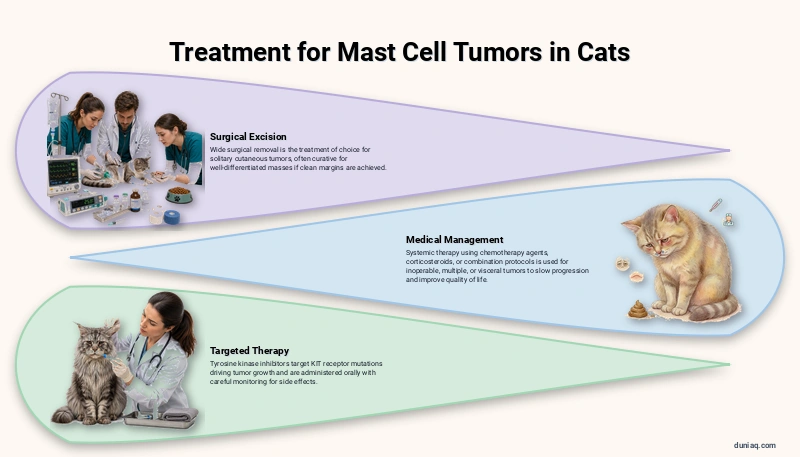
Surgical Excision
Wide surgical removal is the treatment of choice for solitary cutaneous mast cell tumors. The surgeon excises the mass with a margin of normal tissue to ensure complete removal. For well‑differentiated skin tumors, this procedure is often curative. If histopathology reveals incomplete margins, a second surgery or additional local therapy may be recommended to prevent recurrence.
Medical Management
For cats with inoperable tumors, multiple masses, or visceral involvement, systemic therapy becomes necessary. Chemotherapy agents such as lomustine (CCNU) or vinblastine are used to control mast cell proliferation. Prednisolone, a corticosteroid, can reduce inflammation and directly suppress mast cell growth. The combination of prednisolone with chlorambucil is a common first‑line protocol. Treatment aims to improve quality of life and slow progression.
Targeted Therapy
Tyrosine kinase inhibitors (TKIs) like toceranib phosphate (Palladia) and masitinib target KIT receptor mutations driving tumor growth. Although KIT mutations are less common in feline MCTs than in dogs, TKIs have shown benefit in some cats, particularly with gross disease. These medications are administered orally and require careful monitoring for side effects such as gastrointestinal upset or myelosuppression.
- Surgical removal with clean margins is curative for many skin tumors.
- Oral corticosteroids can reduce tumor‑associated swelling and histamine release.
- Chemotherapy is reserved for high‑grade or visceral mast cell tumors.
- Targeted therapy may be considered if the tumor expresses relevant mutations.
- Supportive care includes antihistamines (diphenhydramine) and H2 blockers (famotidine) to manage paraneoplastic histamine effects.
Home Care and Aftercare Instructions

After surgery, the incision site must be kept clean and dry. An Elizabethan collar prevents licking or chewing. Activity restriction is recommended until sutures are removed, typically 10–14 days. Owners should monitor for redness, swelling, or discharge and report any concerns to their veterinarian. Prescribed pain medications and antibiotics must be given as directed and on schedule.
For cats on long‑term medical therapy, consistent administration of oral medications like prednisolone or famotidine is critical. Regular follow‑up visits with physical examinations, blood work, and imaging reassess tumor status. Owners should keep a log of appetite, weight, and any new lumps. Early detection of recurrence or new tumors can be managed more effectively when caught quickly. Any sudden deterioration warrants immediate re‑evaluation.
Recovery Outlook and Monitoring
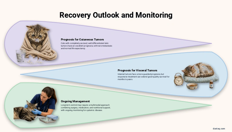
Many cats with cutaneous mast cell tumors have an excellent prognosis after complete surgical excision. Well‑differentiated skin tumors rarely metastasize and do not typically shorten life expectancy. Routine monitoring for new skin masses and periodic veterinary check‑ups are sufficient for these patients. Cats with visceral mast cell tumors face a more guarded prognosis, but responsive treatment can extend good‑quality survival for months to years.
Prognosis depends on tumor location, histological grade, completeness of removal, and response to therapy. Cats that achieve remission with chemotherapy or targeted drugs may enjoy long‑term control. Ongoing management usually involves a multimodal approach combining surgery, medication, and nutritional support. Owners should understand that while a cure is possible for many cutaneous tumors, systemic disease often requires lifelong monitoring and occasional treatment adjustments.
Frequently Asked Questions (FAQ) about Mast Cell Tumors in Cats
Are mast cell tumors in cats always cancerous?
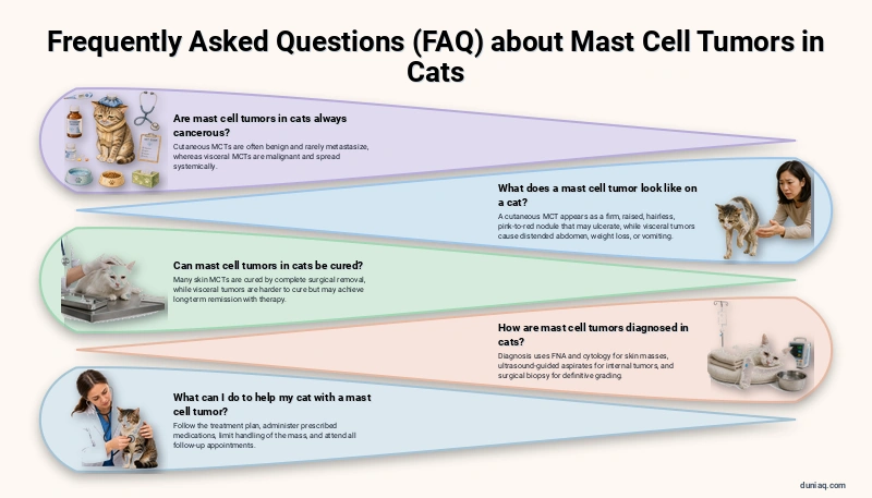
Not all mast cell tumors in cats behave aggressively. Cutaneous mast cell tumors are often benign, particularly the well‑differentiated form common in cats. They rarely metastasize. Visceral mast cell tumors, however, are malignant and spread systemically. The term “tumor” refers to abnormal growth; malignancy is determined by histopathology and staging.
What does a mast cell tumor look like on a cat?
A cutaneous mast cell tumor typically appears as a firm, raised nodule that may be hairless and pink to red. It can fluctuate in size after rubbing and sometimes ulcerates. Multiple lumps can develop. Visceral tumors are not externally visible, but affected cats may have a distended abdomen, weight loss, or vomiting.
Can mast cell tumors in cats be cured?
Yes, many skin mast cell tumors are cured by complete surgical removal. If the tumor is low‑grade and excised with clean margins, recurrence is uncommon. Visceral mast cell tumors are more difficult to cure, but long‑term remission is achievable with chemotherapy or targeted therapy. Each case is evaluated individually by a veterinary oncologist.
How are mast cell tumors diagnosed in cats?
Diagnosis begins with fine‑needle aspiration and cytology of the mass, which reveals mast cells. For internal tumors, ultrasound‑guided aspirates are used. Blood tests and imaging help assess spread. Surgical biopsy with histopathology confirms the tumor type and grade, guiding treatment planning and prognosis.
What can I do to help my cat with a mast cell tumor?
Follow your veterinarian’s treatment plan, which may include surgery, medications, and dietary adjustments. Administer prescribed antihistamines and gastroprotectants to counteract histamine release. Limit handling of the mass to prevent degranulation. Attend all follow‑up appointments and promptly report any new lumps, loss of appetite, or lethargy.
Mast Cell Tumors in Cats Conclusion
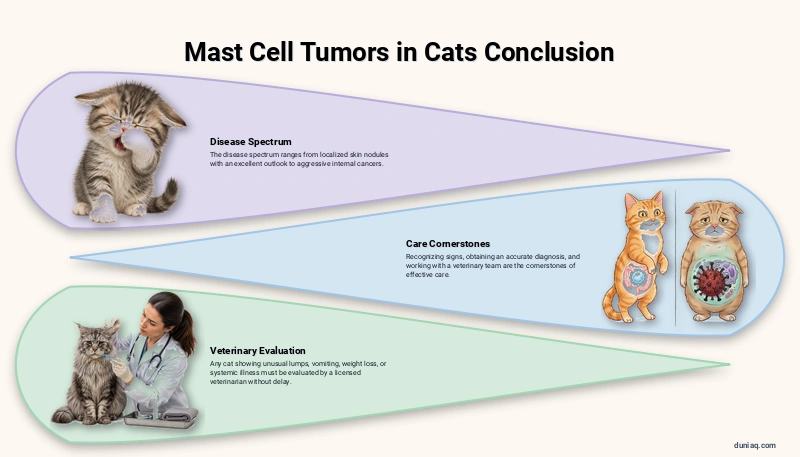
Mast cell tumors in cats encompass a broad spectrum of disease, from localized skin nodules with an excellent outlook to aggressive internal cancers. Recognizing the signs, obtaining an accurate diagnosis, and working with a veterinary team are the cornerstones of effective care. This information is for general educational purposes only. Any cat showing unusual lumps, vomiting, weight loss, or systemic illness must be evaluated by a licensed veterinarian without delay.