Retinal detachment in cats is a serious ophthalmic condition that can lead to partial or complete vision loss if not addressed promptly. The retina, a thin layer of light-sensitive tissue lining the back of the eye, separates from its underlying supportive layers during detachment.
This condition can affect cats of any age, breed, or sex, though certain predisposing factors increase the risk substantially. Understanding the causes, warning signs, and available treatments is essential for cat owners who want to protect their feline companion’s eyesight and overall well-being.
What Is Retinal Detachment in Cats?
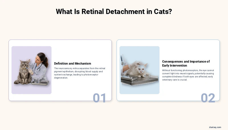
Retinal detachment in cats occurs when the neurosensory retina separates from the retinal pigment epithelium (RPE), the layer responsible for nourishing and supporting retinal cells. This separation disrupts the retina’s blood supply and nutrient exchange, causing progressive degeneration of photoreceptor cells.
Without these functioning photoreceptors, the affected eye cannot properly convert light into neural signals. If both eyes are involved, the cat may experience complete blindness. Early veterinary intervention is critical to preserving any remaining vision and addressing underlying systemic disease.
Types of Retinal Detachment in Cats
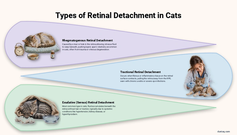
Veterinary ophthalmologists classify retinal detachment in cats into three primary types, each with distinct mechanisms and implications for treatment. Understanding the specific type helps guide the therapeutic approach and determine the prognosis for visual recovery.
Rhegmatogenous Retinal Detachment
This type results from a tear, hole, or break in the retina that allows vitreous fluid to seep beneath the retinal layers, physically pushing them apart. Rhegmatogenous detachments in cats are relatively uncommon compared to other types but may occur following ocular trauma or degenerative changes in the vitreous humor.
Tractional Retinal Detachment
Tractional detachment occurs when fibrous or inflammatory tissue forms on the retinal surface and contracts, mechanically pulling the retina away from the underlying RPE. This type is sometimes seen in cats with chronic intraocular inflammation, also known as uveitis, or following severe eye infections.
Exudative (Serous) Retinal Detachment
Exudative detachment is the most frequently diagnosed type in cats. Fluid accumulates beneath the retina without any tear or traction being present. This fluid buildup typically results from systemic conditions such as hypertension, kidney disease, or hyperthyroidism that damage the blood vessels supplying the retina.
Causes and Risk Factors
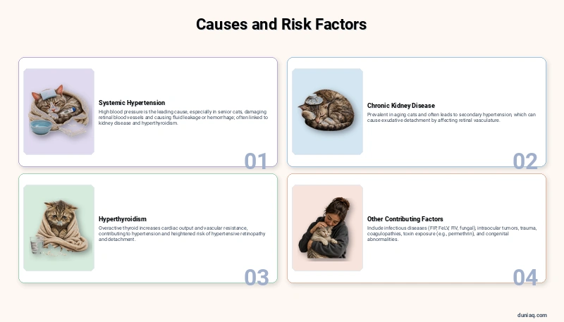
Retinal detachment in cats is often secondary to an underlying systemic disease rather than a primary ocular problem. Identifying and treating the root cause is essential for managing the detachment and preventing recurrence. Several well-documented conditions predispose cats to this serious eye disorder.
Systemic Hypertension
High blood pressure is the leading cause of retinal detachment in cats, particularly in senior felines. Elevated blood pressure damages the delicate retinal blood vessels, causing them to leak fluid or hemorrhage beneath the retina. Hypertension in cats is frequently associated with chronic kidney disease and hyperthyroidism.
Chronic Kidney Disease
Kidney disease is extremely prevalent in aging cats and often leads to secondary hypertension. The kidneys play a central role in regulating blood pressure through the renin-angiotensin-aldosterone system. When kidney function deteriorates, blood pressure may rise unchecked, ultimately affecting the retinal vasculature and causing exudative detachment.
Hyperthyroidism
An overactive thyroid gland increases cardiac output and peripheral vascular resistance, contributing to elevated blood pressure. Cats with untreated or poorly managed hyperthyroidism face a heightened risk of hypertensive retinopathy and subsequent retinal detachment. Regular thyroid screening is advisable for middle-aged and senior cats.
Other Contributing Factors
- Infectious diseases: Feline infectious peritonitis (FIP), feline leukemia virus (FeLV), feline immunodeficiency virus (FIV), and systemic fungal infections can cause uveitis leading to retinal detachment.
- Intraocular neoplasia: Tumors within the eye can physically displace or damage the retina.
- Trauma: Blunt force injury to the head or eye may cause retinal tears or hemorrhage.
- Coagulopathies: Blood clotting disorders can cause retinal hemorrhages and fluid accumulation, as can exposure to certain toxins. Cats accidentally exposed to permethrin-based products may experience severe systemic effects that indirectly compromise ocular health.
- Congenital abnormalities: Rarely, retinal dysplasia or developmental defects may predispose kittens to detachment.
Signs and Symptoms of Retinal Detachment in Cats
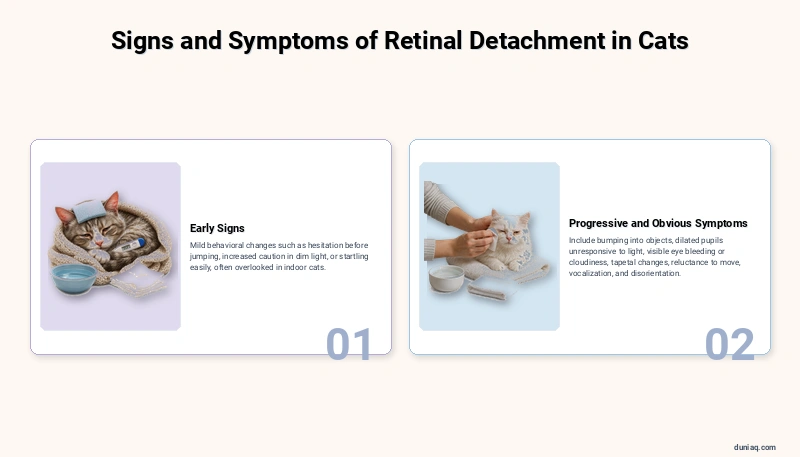
Cats are remarkably adept at compensating for gradual vision loss, which means retinal detachment may progress significantly before an owner notices behavioral changes. Recognizing the subtle and overt signs of this condition can mean the difference between preserving partial vision and permanent blindness.
Early Signs
In the initial stages, cats may show only mild behavioral changes. They might hesitate before jumping onto familiar surfaces, seem slightly more cautious in dim lighting, or startle more easily when approached from certain angles. These early indicators are easy to overlook, especially in indoor cats with predictable routines.
Progressive and Obvious Symptoms
- Bumping into furniture, walls, or objects, particularly in unfamiliar environments
- Dilated pupils (mydriasis) that do not constrict normally in bright light
- Visible bleeding or cloudiness within the eye
- A change in the appearance of the eye’s reflective layer (tapetum) when light shines on it
- Reluctance to move, play, or navigate stairs
- Increased vocalization or signs of anxiety and disorientation
- Walking with a lowered head or whiskers pushed forward to navigate by touch
Some of these ocular symptoms may resemble other eye conditions. For instance, redness and discharge could also suggest pink eye in cats, which is a different condition with distinct causes and treatment. A veterinary examination is necessary to differentiate between these conditions accurately.
When to Seek Emergency Veterinary Care
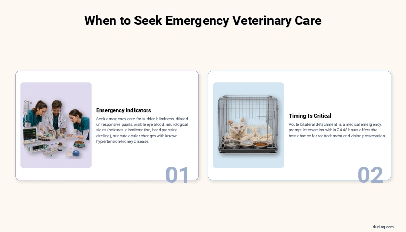
Retinal detachment in cats should always be treated as urgent. However, certain presentations demand immediate emergency attention to maximize the chance of preserving vision and addressing life-threatening underlying conditions.
Seek emergency veterinary care if your cat:
- Suddenly becomes completely blind (bumping into everything, unable to find food or water)
- Develops sudden, widely dilated pupils that do not respond to light
- Shows visible blood inside the eye (hyphema)
- Exhibits neurological signs such as seizures, severe disorientation, head pressing, or circling alongside vision changes
- Has known hypertension or kidney disease and develops any acute ocular changes
Acute bilateral retinal detachment is a medical emergency. The longer the retina remains detached, the greater the photoreceptor damage becomes, and the less likely vision recovery will be. Prompt intervention within the first 24 to 48 hours offers the best chance for reattachment and functional vision preservation.
Diagnosis of Retinal Detachment in Cats
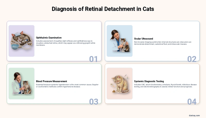
Diagnosing retinal detachment in cats requires a thorough ophthalmic examination combined with systemic diagnostic testing to identify underlying causes. Your veterinarian may refer your cat to a board-certified veterinary ophthalmologist for advanced evaluation and management.
Ophthalmic Examination
The veterinarian will perform a complete eye examination including assessment of pupillary light reflexes, which are often absent or reduced in affected eyes. Direct and indirect ophthalmoscopy allows visualization of the retina itself, where a detached retina may appear as a billowing, grayish-white membrane floating in the vitreous cavity.
Ocular Ultrasound
When the eye’s internal structures cannot be visualized due to hemorrhage, cataracts, or other opacities, ocular ultrasonography is invaluable. This non-invasive imaging technique can clearly demonstrate retinal detachment, identify subretinal fluid, and detect intraocular masses. It is also useful for assessing the extent and type of detachment.
Blood Pressure Measurement
Given that systemic hypertension is the most common cause of retinal detachment in cats, blood pressure measurement is an essential component of the diagnostic workup. Doppler or oscillometric methods are used to obtain readings, and consistently elevated values confirm hypertensive disease requiring treatment.
Systemic Diagnostic Testing
A comprehensive diagnostic panel typically includes:
- Complete blood count (CBC): To assess for infection, anemia, or inflammatory conditions
- Serum biochemistry panel: To evaluate kidney and liver function
- Urinalysis: To further assess renal health and urine concentrating ability
- Thyroid hormone levels (T4): To screen for hyperthyroidism
- Infectious disease testing: FeLV, FIV, FIP, and fungal titers as clinically indicated
- Electroretinography (ERG): To assess retinal function and determine whether the photoreceptors remain viable, which influences prognosis
Differentiating Retinal Detachment From Similar Conditions
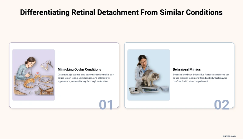
Several ocular and systemic conditions in cats can mimic the signs of retinal detachment, making accurate diagnosis crucial. Cataracts, glaucoma, and severe anterior uveitis can all cause vision loss, changes in pupil size, and alterations in eye appearance. Certain systemic conditions may also cause behavioral changes that mimic blindness.
Stress-related behavioral changes, such as those seen in cats with Pandora syndrome, can sometimes include disorientation or altered activity patterns that might initially be confused with vision impairment. A thorough veterinary evaluation ensures the correct diagnosis and appropriate treatment plan is pursued.
Treatment Options for Retinal Detachment in Cats
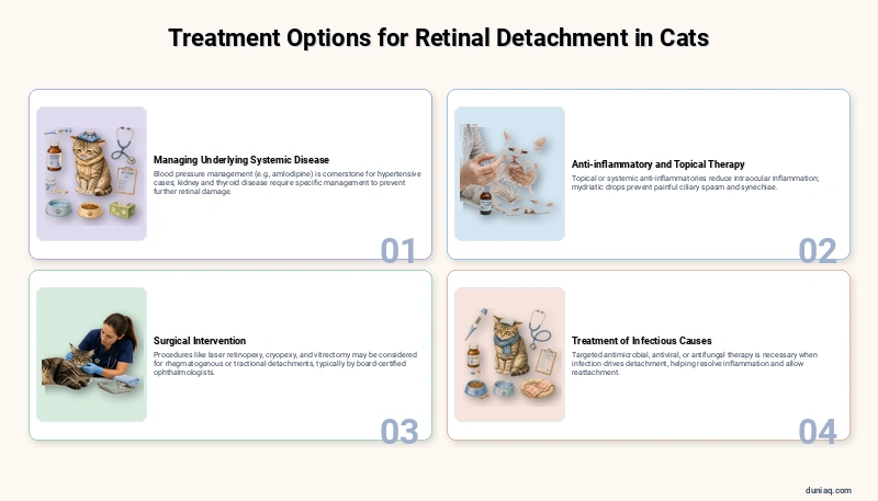
Treatment of retinal detachment in cats focuses on two parallel goals: addressing the underlying systemic cause and managing the detachment itself to maximize the potential for retinal reattachment and vision preservation. Treatment strategies vary based on the type and cause of detachment.
Managing Underlying Systemic Disease
Because exudative retinal detachment secondary to hypertension is the most prevalent form, blood pressure management is often the cornerstone of treatment. Amlodipine besylate is the antihypertensive medication most commonly prescribed for cats with systemic hypertension. Your veterinarian will determine the appropriate medication and dosage based on your cat’s individual needs.
If chronic kidney disease is present, a comprehensive renal management plan including dietary modification, fluid therapy, and phosphorus binders may be recommended. Hyperthyroid cats may require anti-thyroid medication, radioactive iodine therapy, or surgical thyroidectomy. Treating the root cause is fundamental to preventing further retinal damage.
Anti-inflammatory and Topical Therapy
When uveitis accompanies retinal detachment, topical or systemic anti-inflammatory medications may be prescribed to reduce intraocular inflammation. Topical corticosteroids or nonsteroidal anti-inflammatory drops can help manage inflammation within the eye, while systemic medications address broader inflammatory processes. Mydriatic drops may also be used to prevent painful ciliary spasm and synechiae formation.
Surgical Intervention
Surgical treatment for retinal detachment in cats is less commonly performed than in human medicine, but specialized procedures do exist. Laser retinopexy, cryopexy, and vitrectomy may be considered in specific cases, particularly rhegmatogenous or tractional detachments that do not respond to medical management alone.
These advanced surgical procedures are typically performed by board-certified veterinary ophthalmologists at referral centers. The American College of Veterinary Ophthalmologists maintains a directory of specialists who can provide these services. Surgical outcomes depend heavily on the duration of detachment and the viability of the retinal tissue at the time of intervention.
Treatment of Infectious Causes
When retinal detachment results from infectious disease, targeted antimicrobial, antiviral, or antifungal therapy is necessary. Treating the infection helps resolve the inflammatory process driving fluid accumulation or traction, giving the retina an opportunity to reattach once the underlying cause is controlled.
Home Care and Aftercare Following Treatment
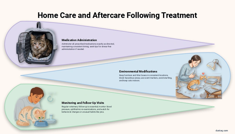
Once your cat has received initial treatment for retinal detachment, ongoing home care and monitoring play a vital role in recovery. Close collaboration with your veterinarian ensures the best possible outcome for your cat’s vision and overall health.
Medication Administration
Administer all prescribed medications exactly as directed by your veterinarian. This may include oral antihypertensive medication, topical eye drops, or systemic anti-inflammatory drugs. Maintain consistent timing for each medication dose. If your cat is difficult to medicate, ask your veterinarian for tips on safe and stress-free administration techniques.
Environmental Modifications
For cats with impaired or lost vision, making environmental adjustments helps reduce injury risk and anxiety:
- Keep furniture and litter boxes in consistent locations — do not rearrange your home
- Block access to stairs, balconies, and other hazardous areas
- Use scent markers or textured mats near food, water, and litter stations to help your cat navigate
- Avoid startling your cat — speak softly before approaching or touching
- Keep your cat indoors at all times to prevent accidents
Monitoring and Follow-Up Visits
Regular veterinary follow-up is essential. Blood pressure should be monitored frequently after initiating antihypertensive therapy to ensure values remain within the target range. Ophthalmic re-examinations help assess whether the retina is reattaching and whether the treatment plan needs adjustment.
Watch for any changes in your cat’s behavior, appetite, or litter box habits at home, as these may indicate progression of underlying disease. Unusual behaviors such as eating non-food items could suggest pica in cats, a behavioral condition worth reporting to your vet to rule out nutritional deficiencies or stress that may complicate recovery.
Recovery Outlook for Cats With Retinal Detachment
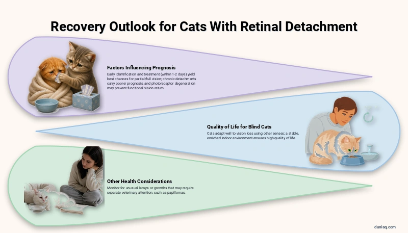
The prognosis for retinal detachment in cats varies significantly depending on several key factors. Cats whose detachments are identified and treated early — particularly within the first one to two days — have the best chance of partial or full vision recovery. Chronic detachments where the retina has been separated for days to weeks carry a much poorer visual prognosis.
The underlying cause also heavily influences outcomes. Cats with well-managed hypertension may experience retinal reattachment and improved vision over weeks to months. However, if photoreceptor degeneration has already occurred, functional vision may not return even after the retina physically reattaches.
Cats that remain permanently blind can still enjoy a high quality of life. Felines rely heavily on their other senses — hearing, whiskers, and scent — and most adapt remarkably well to vision loss in a stable, familiar indoor environment. Owners should focus on providing a safe, enriched, and consistent living space.
It is worth noting that some oral or skin growths may also require veterinary attention during this time. If you notice any unusual lumps, your veterinarian can determine whether they could be something like papillomas in cats or another condition requiring separate treatment.
Prevention Strategies
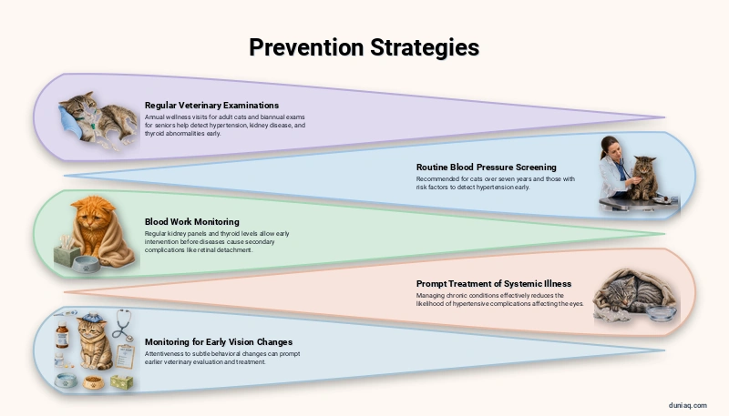
While not all cases of retinal detachment in cats can be prevented, proactive health management significantly reduces the risk. The following strategies are recommended for all cat owners, particularly those with middle-aged and senior felines.
- Regular veterinary examinations: Annual wellness visits for adult cats and biannual exams for seniors help detect hypertension, kidney disease, and thyroid abnormalities early.
- Routine blood pressure screening: The International Society of Feline Medicine recommends routine blood pressure measurement in cats over seven years of age and in any cat with known risk factors.
- Blood work monitoring: Regular kidney panels and thyroid levels allow early intervention before these diseases cause secondary complications like retinal detachment.
- Prompt treatment of systemic illness: Managing chronic conditions effectively reduces the likelihood of hypertensive complications affecting the eyes.
- Monitoring for early vision changes: Being attentive to subtle behavioral changes can prompt earlier veterinary evaluation and treatment.
Frequently Asked Questions (FAQ) About Retinal Detachment in Cats
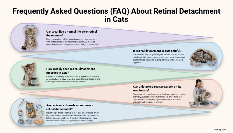
Can a cat live a normal life after retinal detachment?
Many cats adapt exceptionally well to partial or complete vision loss. Cats rely heavily on their other senses, including hearing, touch through their whiskers, and smell. With a stable indoor environment and consistent routines, blind cats can navigate confidently, play, and maintain a high quality of life. The most important factor is managing any underlying disease that caused the detachment.
Is retinal detachment in cats painful?
Retinal detachment itself is generally not considered painful. However, associated conditions such as glaucoma (increased intraocular pressure) or severe uveitis can cause significant ocular discomfort. Signs of eye pain in cats include squinting, excessive tearing, pawing at the eye, and decreased appetite. If you suspect your cat is in pain, prompt veterinary evaluation is needed.
How quickly does retinal detachment progress in cats?
Retinal detachment can occur suddenly, sometimes within hours, particularly in cases of acute hypertensive crisis. In other situations, it may develop gradually over days to weeks. Acute bilateral detachment causing sudden blindness is the presentation that most commonly alerts owners to a problem. The rapid onset underscores why immediate veterinary attention is so critical.
Can a detached retina reattach on its own in cats?
In some cases, particularly when the underlying cause such as hypertension is effectively treated, the subretinal fluid may reabsorb and the retina can reattach without surgical intervention. However, this depends on prompt treatment and the duration of detachment. Spontaneous reattachment without addressing the root cause is unlikely, and delayed treatment reduces the chances of functional vision returning.
Are certain cat breeds more prone to retinal detachment?
Retinal detachment in cats is not strongly breed-specific but is more closely linked to age-related systemic conditions. Senior cats of any breed are at higher risk due to the prevalence of hypertension, chronic kidney disease, and hyperthyroidism in older feline populations. Some breeds predisposed to polycystic kidney disease, such as Persians, may face an indirectly elevated risk through kidney-related hypertension. As noted by Cornell University’s Feline Health Center, regular screening is advisable for all aging cats.
Conclusion
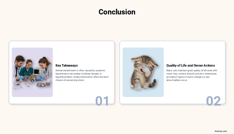
Retinal detachment in cats is a serious condition that requires timely veterinary intervention to offer the best chance of preserving vision. Understanding the causes — most commonly systemic hypertension secondary to kidney disease or hyperthyroidism — empowers cat owners to pursue preventive screening and early treatment.
With proper management, many cats can maintain good quality of life even when vision loss occurs. This article is intended for general informational purposes only. If your cat shows any signs of vision changes or eye abnormalities, please consult a licensed veterinarian promptly for proper diagnosis and treatment.